

IHC-plus(tm) MC5R / MC5 Receptor Antibody (Internal)
LS-A3805
ApplicationsImmunoHistoChemistry, ImmunoHistoChemistry Paraffin
Product group Antibodies
ReactivityHuman
TargetMC5R
Overview
- SupplierLifeSpan BioSciences
- Product NameIHC-plus(tm) MC5R / MC5 Receptor Antibody (Internal)
- Delivery Days Customer23
- Application Supplier NoteImmunohistochemistry: The unconjugated form was validated for use in immunohistochemistry on a panel of 21 formalin-fixed, paraffin-embedded (FFPE) human tissues after proteinase K antigen retrieval. After incubation with the primary antibody, slides were incubated with biotinylated secondary antibody, followed by alkaline phosphatase-streptavidin and chromogen. The stained slides were evaluated by a pathologist to confirm staining specificity. The optimal working concentration for the unconjugated form was determined to be 10 ug/ml.. IHC, IHC-P (10 µg/ml) Immunohistochemistry: The unconjugated form was validated for use in immunohistochemistry on a panel of 21 formalin-fixed, paraffin-embedded (FFPE) human tissues after proteinase K antigen retrieval. After incubation with the primary antibody, slides were incubated with biotinylated secondary antibody, followed by alkaline phosphatase-streptavidin and chromogen. The stained slides were evaluated by a pathologist to confirm staining specificity. The optimal working concentration for the unconjugated form was determined to be 10 ug/ml.
- ApplicationsImmunoHistoChemistry, ImmunoHistoChemistry Paraffin
- CertificationResearch Use Only
- ClonalityPolyclonal
- Concentration1 mg/ml
- ConjugateUnconjugated
- Gene ID4161
- Target nameMC5R
- Target descriptionmelanocortin 5 receptor
- Target synonymsMC2, melanocortin receptor 5
- HostRabbit
- ReactivityHuman
- Storage Instruction-20°C or -80°C,2°C to 8°C
- UNSPSC41116161
Related products
Product group Antibodies
MC5R AntibodyCSB-PA003205
ApplicationsImmunoFluorescence, Western Blot, ELISA
ReactivityHuman, Mouse, Rat
TargetMC5R
- SizePrice
Product group Antibodies
Anti-MC-2 AntibodyA84492
ApplicationsWestern Blot, ELISA, ImmunoHistoChemistry
ReactivityHuman, Mouse
- SizePrice

Product group Antibodies
ApplicationsWestern Blot, ImmunoHistoChemistry
ReactivityHuman
TargetMC5R
- SizePrice
Product group Antibodies

Goat anti-MC5REB08447
ApplicationsWestern Blot, ELISA, ImmunoHistoChemistry
ReactivityHuman, Mouse
TargetMC5R
- SizePrice

Product group Antibodies
Anti-MC5R AntibodyHPA042365
ApplicationsImmunoHistoChemistry
ReactivityHuman
TargetMC5R
- SizePrice
Product group Antibodies

MC5R / MC5 Receptor AntibodyLS-C402434
ApplicationsELISA, ImmunoHistoChemistry
ReactivityHuman
TargetMC5R
- SizePrice
Product group Antibodies
Anti-MC5-R (N-term) Antibody107-10105
ApplicationsWestern Blot
ReactivityHuman
TargetMC5R
- SizePrice
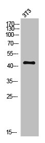
![Various tissue extracts (50 μg) were separated by 10% SDS-PAGE, and the membrane was blotted with MC5R antibody [HL3178] (GTX640696) diluted at 1:1000. The HRP-conjugated anti-rabbit IgG antibody (GTX213110-01) was used to detect the primary antibody, and the signal was developed with Trident ECL plus-Enhanced.](https://www.genetex.com/upload/website/prouct_img/normal/GTX640696/GTX640696_T-45488_20240823_WB_M_tissue_24082802_692.webp)
Product group Antibodies
MC5 Receptor antibody [HL3178]GTX640696
ApplicationsWestern Blot
ReactivityHuman, Mouse
TargetMC5R
- SizePrice
Product group Antibodies
MC5 Receptor Polyclonal AntibodyBS-11418R
ApplicationsImmunoFluorescence, Western Blot, ELISA, ImmunoCytoChemistry, ImmunoHistoChemistry, ImmunoHistoChemistry Frozen, ImmunoHistoChemistry Paraffin
ReactivityHuman, Mouse, Rabbit
TargetMC5R
- SizePrice