

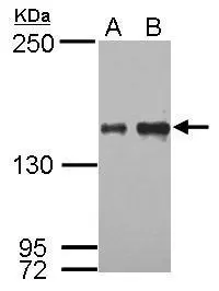
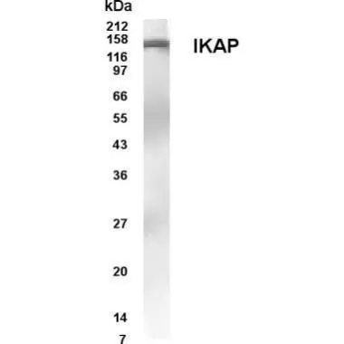
Sample (30 μg of whole cell lysate) A: HeLa B: HepG2 5% SDS PAGE GTX110274 diluted at 1:1000 The HRP-conjugated anti-rabbit IgG antibody (GTX213110-01) was used to detect the primary antibody.
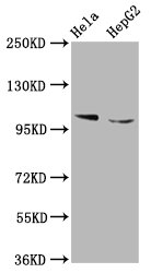
![IKAP antibody [C1C2], Internal detects IKAP protein by western blot analysis. A. 30 μg GL261 whole cell lysate/extract B. 30 μg Raw264.7 whole cell lysate/extract 5% SDS-PAGE IKAP antibody [C1C2], Internal (GTX110274) dilution: 1:500 The HRP-conjugated anti-rabbit IgG antibody (GTX213110-01) was used to detect the primary antibody.](https://www.genetex.com/upload/website/prouct_img/normal/GTX110274/GTX110274_40478_WB_M_w_23060500_958.webp "IKAP antibody [C1C2], Internal detects IKAP protein by western blot analysis. A. 30 μg GL261 whole cell lysate/extract B. 30 μg Raw264.7 whole cell lysate/extract 5% SDS-PAGE IKAP antibody [C1C2], Internal (GTX110274) dilution: 1:500 The HRP-conjugated anti-rabbit IgG antibody (GTX213110-01) was used to detect the primary antibody.")
![IKAP antibody [C1C2], Internal detects IKAP protein at cytoplasm by immunofluorescent analysis. Sample: HeLa cells were fixed in ice-cold MeOH for 5 min. Green: IKAP protein stained by IKBKAP antibody [C1C2], Internal (GTX110274) diluted at 1:500. Blue: Hoechst 33343 staining.](https://www.genetex.com/upload/website/prouct_img/normal/GTX110274/GTX110274_40478_IFA_w_23060500_425.webp "IKAP antibody [C1C2], Internal detects IKAP protein at cytoplasm by immunofluorescent analysis. Sample: HeLa cells were fixed in ice-cold MeOH for 5 min. Green: IKAP protein stained by IKBKAP antibody [C1C2], Internal (GTX110274) diluted at 1:500. Blue: Hoechst 33343 staining.")
Sample (30 μg of whole cell lysate) A: HeLa B: HepG2 5% SDS PAGE GTX110274 diluted at 1:1000 The HRP-conjugated anti-rabbit IgG antibody (GTX213110-01) was used to detect the primary antibody.
IKAP antibody [C1C2], Internal
GTX110274
ApplicationsImmunoFluorescence, Western Blot, ImmunoCytoChemistry
Product group Antibodies
ReactivityHuman, Mouse
TargetELP1
Overview
- SupplierGeneTex
- Product NameIKAP antibody [C1C2], Internal
- Delivery Days Customer9
- Application Supplier NoteWB: 1:500-1:3000. ICC/IF: 1:100-1:1000. *Optimal dilutions/concentrations should be determined by the researcher.Not tested in other applications.
- ApplicationsImmunoFluorescence, Western Blot, ImmunoCytoChemistry
- CertificationResearch Use Only
- ClonalityPolyclonal
- Concentration1 mg/ml
- ConjugateUnconjugated
- Gene ID8518
- Target nameELP1
- Target descriptionelongator acetyltransferase complex subunit 1
- Target synonymsDYS, FD, IKAP, IKBKAP, IKI3, TOT1, elongator complex protein 1, IKK complex-associated protein, ikappaB kinase complex-associated protein, inhibitor of kappa light polypeptide gene enhancer in B-cells, kinase complex-associated protein
- HostRabbit
- IsotypeIgG
- Protein IDO95163
- Protein NameElongator complex protein 1
- Scientific DescriptionThe protein encoded by this gene is a scaffold protein and a regulator for 3 different kinases involved in proinflammatory signaling. This encoded protein can bind NF-kappa-B-inducing kinase (NIK) and IKKs through separate domains and assemble them into an active kinase complex. Mutations in this gene have been associated with familial dysautonomia. [provided by RefSeq]
- ReactivityHuman, Mouse
- Storage Instruction-20°C or -80°C,2°C to 8°C
- UNSPSC41116161
Datasheet
Related products
Product group Antibodies
ELP1 AntibodyCSB-PA011571LA01HU
ApplicationsImmunoFluorescence, ImmunoPrecipitation, Western Blot, ELISA, ImmunoHistoChemistry
ReactivityHuman
TargetELP1
- SizePrice
Product group Antibodies
Anti-IKAP AntibodyA48868
ApplicationsWestern Blot, ELISA, ImmunoCytoChemistry
ReactivityHuman, Mouse
- SizePrice

Product group Antibodies
Anti-IKBKAP ELP1 AntibodyA31687-1
ApplicationsWestern Blot, ImmunoHistoChemistry
ReactivityHuman, Mouse, Rat
TargetELP1
- SizePrice
Product group Antibodies

IKBKAP / IKAP AntibodyLS-C831621
ApplicationsELISA, ImmunoHistoChemistry
ReactivityHuman
TargetELP1
- SizePrice

Product group Antibodies
Anti-IKBKAP AntibodyHPA050686
ApplicationsImmunoHistoChemistry
ReactivityHuman
TargetELP1
- SizePrice
Product group Antibodies
IKAP antibodyGTX30260
ApplicationsWestern Blot
ReactivityHuman
TargetELP1
- SizePrice
Product group Antibodies
ELP1 Polyclonal AntibodyCAC13087
ApplicationsImmunoFluorescence, ImmunoPrecipitation, Western Blot, ELISA, ImmunoHistoChemistry
TargetELP1
- SizePrice
Product group Antibodies
IKAP antibodyGTX64808
ApplicationsWestern Blot, ImmunoHistoChemistry, ImmunoHistoChemistry Paraffin
ReactivityHuman, Mouse, Rat
TargetELP1
- SizePrice
Product group Antibodies
IKAP antibodyGTX70364
ApplicationsWestern Blot, ELISA
ReactivityHuman
- SizePrice