

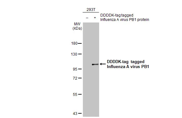
Non-transfected (–) and transfected (+) 293T whole cell extracts (30 μg) were separated by 7.5% SDS-PAGE, and the membrane was blotted with Influenza A virus PB1 protein antibody [HL1715] (GTX637314) diluted at 1:5000. The HRP-conjugated anti-rabbit IgG antibody (GTX213110-01) was used to detect the primary antibody.
![Influenza A (H1N1) viral lysate (0.5 μg) was separated by 7.5% SDS-PAGE, and the membrane was blotted with Influenza A virus PB1 protein antibody [HL1715] (GTX637314) diluted at 1:5000. The HRP-conjugated anti-rabbit IgG antibody (GTX213110-01) was used to detect the primary antibody.](https://www.genetex.com/upload/website/prouct_img/normal/GTX637314/GTX637314_44844_20221028_WB_H1N1_22110201_357.webp "Influenza A (H1N1) viral lysate (0.5 μg) was separated by 7.5% SDS-PAGE, and the membrane was blotted with Influenza A virus PB1 protein antibody [HL1715] (GTX637314) diluted at 1:5000. The HRP-conjugated anti-rabbit IgG antibody (GTX213110-01) was used to detect the primary antibody.")
![Influenza A virus PB1 protein antibody [HL1715] detects Influenza A virus PB1 protein protein by immunohistochemical analysis. Sample:Paraffin-embedded mock and Influenza A virus PB1 protein transfected 293T. Green: Influenza A virus PB1 protein stained by Influenza A virus PB1 protein antibody [HL1715] (GTX637314) diluted at 1:1000. Blue: Fluoroshield with DAPI (GTX30920). Antigen Retrieval: Citrate buffer, pH 6.0, 15 min](https://www.genetex.com/upload/website/prouct_img/normal/GTX637314/GTX637314_44844_20230324_IHC-P_cell_pellet_23032819_115.webp "Influenza A virus PB1 protein antibody [HL1715] detects Influenza A virus PB1 protein protein by immunohistochemical analysis. Sample:Paraffin-embedded mock and Influenza A virus PB1 protein transfected 293T. Green: Influenza A virus PB1 protein stained by Influenza A virus PB1 protein antibody [HL1715] (GTX637314) diluted at 1:1000. Blue: Fluoroshield with DAPI (GTX30920). Antigen Retrieval: Citrate buffer, pH 6.0, 15 min")
Non-transfected (–) and transfected (+) 293T whole cell extracts (30 μg) were separated by 7.5% SDS-PAGE, and the membrane was blotted with Influenza A virus PB1 protein antibody [HL1715] (GTX637314) diluted at 1:5000. The HRP-conjugated anti-rabbit IgG antibody (GTX213110-01) was used to detect the primary antibody.
Influenza A virus PB1 protein antibody [HL1715]
GTX637314
ApplicationsWestern Blot, ImmunoHistoChemistry
Product group Antibodies
ReactivityVirus
Overview
- SupplierGeneTex
- Product NameInfluenza A virus PB1 protein antibody [HL1715]
- Delivery Days Customer9
- Application Supplier NoteWB: 1:1000-1:10000. *Optimal dilutions/concentrations should be determined by the researcher.Not tested in other applications.
- ApplicationsWestern Blot, ImmunoHistoChemistry
- CertificationResearch Use Only
- ClonalityMonoclonal
- Clone IDHL1715
- Concentration1 mg/ml
- ConjugateUnconjugated
- HostRabbit
- IsotypeIgG
- ReactivityVirus
- Storage Instruction-20°C or -80°C,2°C to 8°C
- UNSPSC12352203