

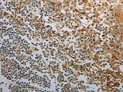
Immunohistochemistry of paraffin-embedded Human tonsil tissue using INHA Polyclonal Antibody at dilution 1:120
Immunohistochemistry of paraffin-embedded Human tonsil tissue using INHA Polyclonal Antibody at dilution 1:120
INHA Polyclonal Antibody
E-AB-10398
ApplicationsImmunoHistoChemistry
Product group Antibodies
TargetINHA
Overview
- SupplierElabscience
- Product NameINHA Polyclonal Antibody
- Delivery Days Customer12
- ApplicationsImmunoHistoChemistry
- Applications SupplierELISA IHC
- CertificationResearch Use Only
- ClonalityPolyclonal
- Concentration1 mg/ml
- ConjugateUnconjugated
- Gene ID3623
- Target nameINHA
- Target descriptioninhibin subunit alpha
- Target synonymsinhibin alpha chain, A-inhibin subunit, inhibin alpha subunit
- HostRabbit
- IsotypeIgG
- Protein IDP05111
- Protein NameInhibin alpha chain
- Scientific DescriptionThis gene encodes the alpha subunit of inhibins A and B protein complexes. These complexes negatively regulate follicle stimulating hormone secretion from the pituitary gland. Inhibins have also been implicated in regulating numerous cellular processes including cell proliferation, apoptosis, immune response and hormone secretion.[
- Storage Instruction-20°C
- UNSPSC41116161
MSDS
Related products
Product group Antibodies

Anti-INHibin-alpha [15E11D5E11]AB03405-1.1-BT
ApplicationsImmunoHistoChemistry
ReactivityHuman
TargetINHA
- SizePrice

Product group Antibodies
Anti-INHA AntibodyA29073
ApplicationsWestern Blot
ReactivityHuman, Mouse, Rat
- SizePrice

Product group Antibodies
Anti-Inhibin alpha/INHA Antibody Picoband(r)A02413-3-CARRIER-FREE
ApplicationsFlow Cytometry, ImmunoFluorescence, Western Blot, ELISA, ImmunoCytoChemistry, ImmunoHistoChemistry
ReactivityHuman, Mouse, Rat
TargetINHA
- SizePrice
Product group Antibodies

ApplicationsWestern Blot, ELISA
ReactivityHuman
TargetINHA
- SizePrice
Product group Antibodies

ApplicationsELISA
ReactivityBovine, Human
TargetINHA
- SizePrice

Product group Antibodies
Anti-INHA AntibodyHPA019141
ApplicationsImmunoCytoChemistry, ImmunoHistoChemistry
ReactivityHuman
TargetINHA
- SizePrice
Product group Antibodies
INHA AntibodyCSB-PA033184
ApplicationsELISA, ImmunoHistoChemistry
ReactivityHuman, Mouse, Rat
TargetINHA
- SizePrice
Product group Antibodies
ApplicationsFlow Cytometry
TargetINHA
- SizePrice
Product group Antibodies
Inhibin alpha Recombinant AntibodyBSM-60582R
ApplicationsImmunoFluorescence, Western Blot, ImmunoCytoChemistry, ImmunoHistoChemistry, ImmunoHistoChemistry Frozen, ImmunoHistoChemistry Paraffin
ReactivityHuman
TargetINHA
- SizePrice