

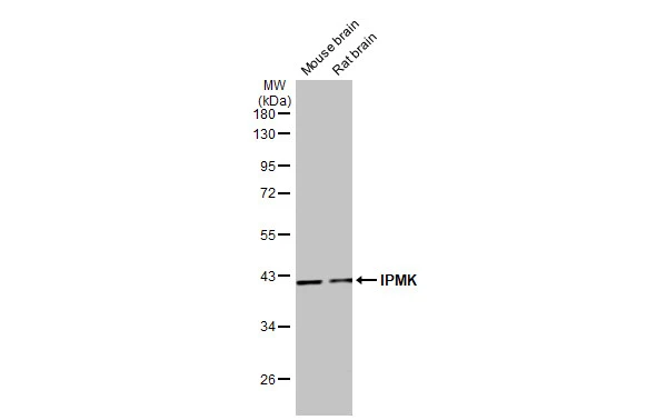
Various tissue extracts (50 μg) were separated by 10% SDS-PAGE, and the membrane was blotted with IPMK antibody [HL2244] (GTX638293) diluted at 1:4000. The HRP-conjugated anti-rabbit IgG antibody (GTX213110-01) was used to detect the primary antibody.
![Non-transfected (–) and transfected (+) A431 whole cell extracts (30 μg) were separated by 10% SDS-PAGE, and the membrane was blotted with IPMK antibody [HL2244] (GTX638293) diluted at 1:4000. The HRP-conjugated anti-rabbit IgG antibody (GTX213110-01) was used to detect the primary antibody.](https://www.genetex.com/upload/website/prouct_img/normal/GTX638293/GTX638293_T-44960_20230317_WB_shRNA_watermark_23032022_724.webp "Non-transfected (–) and transfected (+) A431 whole cell extracts (30 μg) were separated by 10% SDS-PAGE, and the membrane was blotted with IPMK antibody [HL2244] (GTX638293) diluted at 1:4000. The HRP-conjugated anti-rabbit IgG antibody (GTX213110-01) was used to detect the primary antibody.")
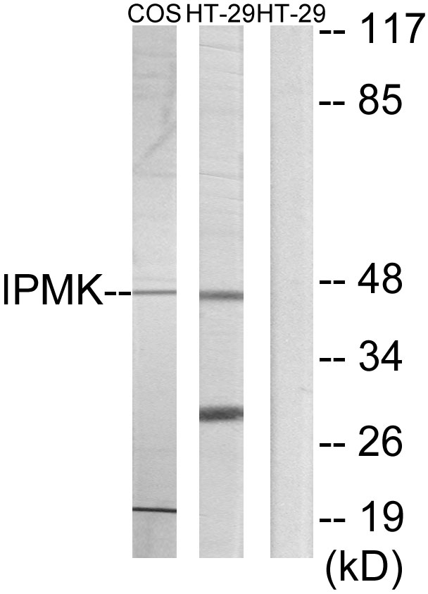
![Various whole cell extracts (30 μg) were separated by 10% SDS-PAGE, and the membrane was blotted with IPMK antibody [HL2244] (GTX638293) diluted at 1:5000. The HRP-conjugated anti-rabbit IgG antibody (GTX213110-01) was used to detect the primary antibody, and the signal was developed with Trident ECL plus-Enhanced.](https://www.genetex.com/upload/website/prouct_img/normal/GTX638293/GTX638293_45026_20230428_WB_23050223_673.webp "Various whole cell extracts (30 μg) were separated by 10% SDS-PAGE, and the membrane was blotted with IPMK antibody [HL2244] (GTX638293) diluted at 1:5000. The HRP-conjugated anti-rabbit IgG antibody (GTX213110-01) was used to detect the primary antibody, and the signal was developed with Trident ECL plus-Enhanced.")
![Non-transfected (–) and transfected (+) 293T whole cell extracts (30 μg) were separated by 10% SDS-PAGE, and the membrane was blotted with IPMK antibody [HL2244] (GTX638293) diluted at 1:500000. The HRP-conjugated anti-rabbit IgG antibody (GTX213110-01) was used to detect the primary antibody.](https://www.genetex.com/upload/website/prouct_img/normal/GTX638293/GTX638293_45026_20230728_WB_B_23073119_550.webp "Non-transfected (–) and transfected (+) 293T whole cell extracts (30 μg) were separated by 10% SDS-PAGE, and the membrane was blotted with IPMK antibody [HL2244] (GTX638293) diluted at 1:500000. The HRP-conjugated anti-rabbit IgG antibody (GTX213110-01) was used to detect the primary antibody.")
Various tissue extracts (50 μg) were separated by 10% SDS-PAGE, and the membrane was blotted with IPMK antibody [HL2244] (GTX638293) diluted at 1:4000. The HRP-conjugated anti-rabbit IgG antibody (GTX213110-01) was used to detect the primary antibody.
IPMK antibody [HL2244]
GTX638293
ApplicationsWestern Blot
Product group Antibodies
ReactivityHuman, Mouse, Rat
TargetIPMK
Overview
- SupplierGeneTex
- Product NameIPMK antibody [HL2244]
- Delivery Days Customer9
- Application Supplier NoteWB: 1:1000-1:10000. *Optimal dilutions/concentrations should be determined by the researcher.Not tested in other applications.
- ApplicationsWestern Blot
- CertificationResearch Use Only
- ClonalityMonoclonal
- Clone IDHL2244
- Concentration2 mg/ml
- ConjugateUnconjugated
- Gene ID253430
- Target nameIPMK
- Target descriptioninositol polyphosphate multikinase
- Target synonymsinositol polyphosphate multikinase, inositol 1,3,4,6-tetrakisphosphate 5-kinase
- HostRabbit
- IsotypeIgG
- Protein IDQ8NFU5
- Protein NameInositol polyphosphate multikinase
- Scientific DescriptionThis gene encodes a member of the inositol phosphokinase family. The encoded protein has 3-kinase, 5-kinase and 6-kinase activities on phosphorylated inositol substrates. The encoded protein plays an important role in the biosynthesis of inositol 1,3,4,5,6-pentakisphosphate, and has a preferred 5-kinase activity. This gene may play a role in nuclear mRNA export. Pseudogenes of this gene are located on the long arm of chromosome 13 and the short arm of chromosome 19. [provided by RefSeq, Dec 2010]
- ReactivityHuman, Mouse, Rat
- Storage Instruction-20°C or -80°C,2°C to 8°C
- UNSPSC41116161
Datasheet
Related products
Product group Antibodies
Anti-IPMK AntibodyA98081
ApplicationsWestern Blot, ELISA
ReactivityHuman, Mouse, Rat
- SizePrice

Product group Antibodies
Anti-IPMK AntibodyA06955-1
ApplicationsImmunoFluorescence, Western Blot, ELISA, ImmunoHistoChemistry
ReactivityHuman, Monkey, Mouse, Rat
TargetIPMK
- SizePrice
Product group Antibodies
Anti-IPMK Antibody107-10502
ApplicationsImmunoFluorescence, Western Blot, ImmunoCytoChemistry
ReactivityHuman, Mouse
TargetIPMK
- SizePrice
Product group Antibodies
IPMK Polyclonal AntibodyBS-12385R
ApplicationsImmunoFluorescence, Western Blot, ELISA, ImmunoCytoChemistry, ImmunoHistoChemistry, ImmunoHistoChemistry Frozen, ImmunoHistoChemistry Paraffin
ReactivityBovine, Equine, Human, Mouse, Rabbit, Rat, Sheep
TargetIPMK
- SizePrice
Product group Antibodies
IPMK AntibodyCSB-PA003058
ApplicationsWestern Blot, ELISA, ImmunoHistoChemistry
ReactivityHuman, Monkey, Mouse, Rat
TargetIPMK
- SizePrice
Product group Antibodies

IPMK Antibody (280-360 aa, C-terminal)LS-C384167
ApplicationsWestern Blot, ELISA, ImmunoHistoChemistry
ReactivityHuman, Monkey, Mouse, Rat
TargetIPMK
- SizePrice
![IPMK antibody [N1C2] detects IPMK protein at cytoplasm and nucleus by immunohistochemical analysis. Sample: Paraffin-embedded mouse intestine. IPMK stained by IPMK antibody [N1C2] (GTX104954) diluted at 1:500. Antigen Retrieval: Citrate buffer, pH 6.0, 15 min](https://www.genetex.com/upload/website/prouct_img/normal/GTX104954/GTX104954_44601_20220527_IHC-P_M_22062121_687.webp)
Product group Antibodies
IPMK antibody [N1C2]GTX104954
ApplicationsImmunoFluorescence, Western Blot, ImmunoCytoChemistry, ImmunoHistoChemistry, ImmunoHistoChemistry Paraffin
ReactivityHuman, Mouse
TargetIPMK
- SizePrice

Product group Antibodies
Anti-IPMK AntibodyHPA037837
ApplicationsImmunoCytoChemistry
ReactivityHuman
TargetIPMK
- SizePrice
Product group Antibodies
IPMK AntibodyPACO00979
ApplicationsWestern Blot, ELISA, ImmunoHistoChemistry
ReactivityHuman, Monkey, Mouse, Rat
TargetIPMK
- SizePrice