

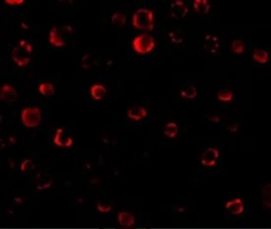
ICC/IF analysis of HeLa cells using GTX31253 IRAK1 antibody. Working concentration : 20 μg/ml

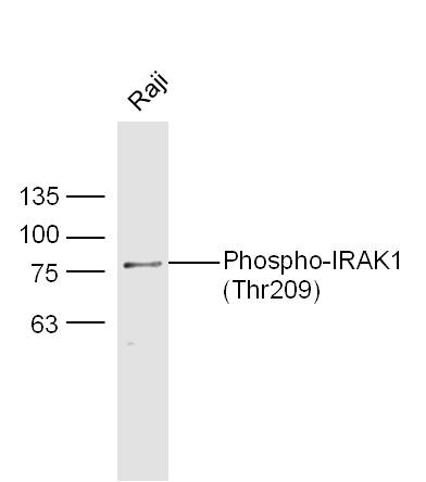
 and HeLa (HL) whole cell lysates using GTX31253 IRAK1 antibody. Working concentration : 1 μg/ml")
ICC/IF analysis of HeLa cells using GTX31253 IRAK1 antibody. Working concentration : 20 μg/ml
IRAK1 antibody
GTX31253
ApplicationsImmunoFluorescence, ImmunoPrecipitation, Western Blot, ELISA, ImmunoCytoChemistry
Product group Antibodies
ReactivityHuman, Mouse, Rat
TargetIRAK1
Overview
- SupplierGeneTex
- Product NameIRAK1 antibody
- Delivery Days Customer9
- Application Supplier NoteWB: 1 microg/mL. ICC/IF: 10 microg/mL. IP: 2 - 4 microg. *Optimal dilutions/concentrations should be determined by the researcher.Not tested in other applications.
- ApplicationsImmunoFluorescence, ImmunoPrecipitation, Western Blot, ELISA, ImmunoCytoChemistry
- CertificationResearch Use Only
- ClonalityPolyclonal
- Concentration1 mg/ml
- ConjugateUnconjugated
- Gene ID3654
- Target nameIRAK1
- Target descriptioninterleukin 1 receptor associated kinase 1
- Target synonymsIRAK, pelle, interleukin-1 receptor-associated kinase 1, Pelle homolog
- HostRabbit
- IsotypeIgG
- Protein IDP51617
- Protein NameInterleukin-1 receptor-associated kinase 1
- Scientific DescriptionThis gene encodes the interleukin-1 receptor-associated kinase 1, one of two putative serine/threonine kinases that become associated with the interleukin-1 receptor (IL1R) upon stimulation. This gene is partially responsible for IL1-induced upregulation of the transcription factor NF-kappa B. Alternatively spliced transcript variants encoding different isoforms have been found for this gene. [provided by RefSeq, Jul 2008]
- ReactivityHuman, Mouse, Rat
- Storage Instruction-20°C or -80°C,2°C to 8°C
- UNSPSC41116161
References
- MicroRNA-146b-5p Suppresses Pro-Inflammatory Mediator Synthesis via Targeting TRAF6, IRAK1, and RELA in Lipopolysaccharide-Stimulated Human Dental Pulp Cells.Read this paper
Datasheet
Related products
Product group Antibodies

IRAK1 (Phospho-Ser376) AntibodyABX012604
ApplicationsELISA, ImmunoHistoChemistry
- SizePrice

Product group Antibodies
Anti-IRAK1 AntibodyA100667
ApplicationsELISA, ImmunoHistoChemistry
ReactivityHuman
- SizePrice

Product group Antibodies
Anti-IRAK-1/IRAK1 Antibody Picoband(r)A01021-1-CARRIER-FREE
ApplicationsFlow Cytometry, ImmunoFluorescence, Western Blot, ImmunoCytoChemistry, ImmunoHistoChemistry
ReactivityHuman, Mouse, Rat
TargetIRAK1
- SizePrice
Product group Antibodies
Anti-IRAK1 Antibody144-12624
ApplicationsImmunoFluorescence, Western Blot
ReactivityHuman, Mouse, Rat
TargetIRAK1
- SizePrice
Product group Antibodies

IRAK1 / IRAK AntibodyLS-C747718
ApplicationsImmunoFluorescence, Western Blot
ReactivityHuman, Mouse, Rat
TargetIRAK1
- SizePrice
Product group Antibodies
ApplicationsFlow Cytometry, ImmunoFluorescence, Western Blot, ImmunoCytoChemistry, ImmunoHistoChemistry, ImmunoHistoChemistry Frozen, ImmunoHistoChemistry Paraffin
ReactivityHuman, Mouse, Rat
TargetIRAK1
- SizePrice
Product group Antibodies
IRAK1 AntibodyCSB-PA003061
ApplicationsImmunoFluorescence, Western Blot, ELISA
ReactivityHuman, Mouse, Rat
TargetIRAK1
- SizePrice
Product group Antibodies
ApplicationsImmunoPrecipitation, Western Blot, ImmunoCytoChemistry, ImmunoHistoChemistry
ReactivityMouse, Rat
TargetIRAK1
- SizePrice
![WB analysis of mouse liver tissue lysate using GTX17299 IRAK1 antibody [8F1A7]. Working concentration : (A) 1 and (B) 2 μg/ml](https://www.genetex.com/upload/website/prouct_img/normal/GTX17299/GTX17299_2437_WB_20180221_w_23060620_282.webp)
Product group Antibodies
IRAK1 antibody [8F1A7]GTX17299
ApplicationsWestern Blot, ELISA, ImmunoHistoChemistry, ImmunoHistoChemistry Paraffin
ReactivityHuman, Mouse, Rat
TargetIRAK1
- SizePrice
Product group Antibodies
IRAK1 antibodyGTX20238
ApplicationsImmunoPrecipitation, Western Blot
ReactivityHuman
TargetIRAK1
- SizePrice