

Immunohistochemical analysis of paraffin-embedded A549 xenograft, using ISCU(GTX115709) antibody at 1:500 dilution.
Antigen Retrieval: Trilogy? (EDTA based, pH 8.0) buffer, 15min
![ISCU antibody [N1C3] detects ISCU protein at mitochondria by immunofluorescent analysis. Sample: HeLa cells were fixed in ice-cold MeOH for 5 min. Green: ISCU stained by ISCU antibody [N1C3] (GTX115709) diluted at 1:500. Blue: Hoechst 33342 staining.](https://www.genetex.com/upload/website/prouct_img/normal/GTX115709/GTX115709_43104_20180307_ICC_IF_w_23060519_116.webp "ISCU antibody [N1C3] detects ISCU protein at mitochondria by immunofluorescent analysis. Sample: HeLa cells were fixed in ice-cold MeOH for 5 min. Green: ISCU stained by ISCU antibody [N1C3] (GTX115709) diluted at 1:500. Blue: Hoechst 33342 staining.")
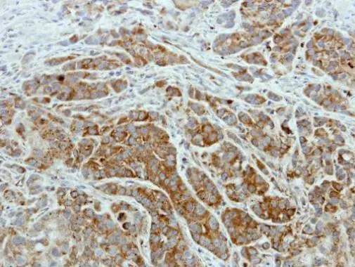
![ISCU antibody [N1C3] detects ISCU protein at cytosol on mouse muscle by immunohistochemical analysis. Sample: Paraffin-embedded mouse muscle. ISCU antibody [N1C3] (GTX115709) dilution: 1:500.
Antigen Retrieval: Trilogy? (EDTA based, pH 8.0) buffer, 15min](https://www.genetex.com/upload/website/prouct_img/normal/GTX115709/GTX115709_40296_IHC_M_w_23060519_174.webp "ISCU antibody [N1C3] detects ISCU protein at cytosol on mouse muscle by immunohistochemical analysis. Sample: Paraffin-embedded mouse muscle. ISCU antibody [N1C3] (GTX115709) dilution: 1:500.
Antigen Retrieval: Trilogy? (EDTA based, pH 8.0) buffer, 15min")
![Non-transfected (–) and transfected (+) 293T whole cell extracts (30 μg) were separated by 15% SDS-PAGE, and the membrane was blotted with ISCU antibody [N1C3] (GTX115709) diluted at 1:5000. The HRP-conjugated anti-rabbit IgG antibody (GTX213110-01) was used to detect the primary antibody.](https://www.genetex.com/upload/website/prouct_img/normal/GTX115709/GTX115709_43104_20180216_WB_B_w_23060519_126.webp "Non-transfected (–) and transfected (+) 293T whole cell extracts (30 μg) were separated by 15% SDS-PAGE, and the membrane was blotted with ISCU antibody [N1C3] (GTX115709) diluted at 1:5000. The HRP-conjugated anti-rabbit IgG antibody (GTX213110-01) was used to detect the primary antibody.")
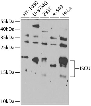
![Various whole cell extracts (30 μg) were separated by 15% SDS-PAGE, and the membrane was blotted with ISCU antibody [N1C3] (GTX115709) diluted at 1:500. The HRP-conjugated anti-rabbit IgG antibody (GTX213110-01) was used to detect the primary antibody.](https://www.genetex.com/upload/website/prouct_img/normal/GTX115709/GTX115709_43104_20180216_WB_w_23060519_869.webp "Various whole cell extracts (30 μg) were separated by 15% SDS-PAGE, and the membrane was blotted with ISCU antibody [N1C3] (GTX115709) diluted at 1:500. The HRP-conjugated anti-rabbit IgG antibody (GTX213110-01) was used to detect the primary antibody.")
Immunohistochemical analysis of paraffin-embedded A549 xenograft, using ISCU(GTX115709) antibody at 1:500 dilution.
Antigen Retrieval: Trilogy? (EDTA based, pH 8.0) buffer, 15min
ISCU antibody [N1C3]
GTX115709
ApplicationsImmunoFluorescence, Western Blot, ImmunoCytoChemistry, ImmunoHistoChemistry, ImmunoHistoChemistry Paraffin
Product group Antibodies
ReactivityHuman, Mouse
TargetISCU
Overview
- SupplierGeneTex
- Product NameISCU antibody [N1C3]
- Delivery Days Customer9
- Application Supplier NoteWB: 1:500-1:10000. ICC/IF: 1:100-1:1000. IHC-P: 1:100-1:1000. *Optimal dilutions/concentrations should be determined by the researcher.Not tested in other applications.
- ApplicationsImmunoFluorescence, Western Blot, ImmunoCytoChemistry, ImmunoHistoChemistry, ImmunoHistoChemistry Paraffin
- CertificationResearch Use Only
- ClonalityPolyclonal
- Concentration0.66 mg/ml
- ConjugateUnconjugated
- Gene ID23479
- Target nameISCU
- Target descriptioniron-sulfur cluster assembly enzyme
- Target synonyms2310020H20Rik, HML, ISU2, NIFU, NIFUN, hnifU, iron-sulfur cluster assembly enzyme ISCU, IscU iron-sulfur cluster scaffold homolog, iron-sulfur cluster assembly enzyme ISCU, mitochondrial, nifU-like N-terminal domain-containing protein
- HostRabbit
- IsotypeIgG
- Protein IDQ9H1K1
- Protein NameIron-sulfur cluster assembly enzyme ISCU
- Scientific DescriptionIron-sulfur (Fe-S) clusters are necessary for several mitochondrial enzymes and other subcellular compartment proteins. They contain sulfur and iron, and are created via several steps that include cysteine desulfurases, iron donors, chaperones, and scaffold proteins. This gene encodes the two isomeric forms, ISCU1 and ISCU2, of the Fe-S cluster scaffold protein. Mutations in this gene have been found in patients with myopathy with severe exercise intolerance and myoglobinuria. [provided by RefSeq]
- ReactivityHuman, Mouse
- Storage Instruction-20°C or -80°C,2°C to 8°C
- UNSPSC41116161
Datasheet
Related products
Product group Antibodies

ISCU AntibodyLS-C830568
ApplicationsWestern Blot, ELISA
ReactivityHuman, Mouse
TargetISCU
- SizePrice

Product group Antibodies
Anti-ISCU AntibodyHPA038602
ApplicationsImmunoCytoChemistry, ImmunoHistoChemistry
ReactivityHuman
TargetISCU
- SizePrice
Product group Antibodies
ISCU AntibodyCSB-PA887955DA01HU
ApplicationsELISA
ReactivityHuman
TargetISCU
- SizePrice

Product group Antibodies
ISCU antibodyGTX66491
ApplicationsWestern Blot
ReactivityHuman
TargetISCU
- SizePrice
Product group Antibodies
Anti-ISCUY058899
ApplicationsWestern Blot, ELISA, ImmunoHistoChemistry
ReactivityHuman
- SizePrice
Product group Antibodies
Anti-ISCU Antibody144-07082
ApplicationsWestern Blot
ReactivityHuman, Mouse, Rat
TargetISCU
- SizePrice