

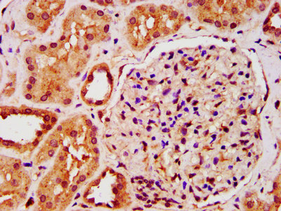
IST1 Antibody, Biotin conjugated
MBS7107942
Product group Antibodies
Overview
- SupplierMyBioSource
- Product NameIST1 Antibody, Biotin conjugated
- Delivery Days Customer16
- CertificationResearch Use Only
- Estimated Purity>95%, Protein G purified
- IsotypeIgG
- Storage Instruction-20°C or -80°C
- UNSPSC12352203
Related products

Product group Antibodies
Anti-hIST1/IST1 Antibody Picoband(r)A05507-2-CARRIER-FREE
ApplicationsFlow Cytometry, ImmunoFluorescence, Western Blot, ELISA, ImmunoCytoChemistry, ImmunoHistoChemistry
TargetIST1
- SizePrice

Product group Antibodies
Anti-IST1 AntibodyHPA054532
ApplicationsWestern Blot, ImmunoCytoChemistry, ImmunoHistoChemistry
TargetIST1
- SizePrice
Product group Antibodies
IST1 AntibodyCSB-PA012171LA01HU
ApplicationsImmunoFluorescence, ELISA, ImmunoHistoChemistry
TargetIST1
- SizePrice
Product group Antibodies
ApplicationsImmunoPrecipitation, Western Blot
TargetIST1
- SizePrice
Product group Antibodies

IST1 AntibodyLS-C830187
ApplicationsWestern Blot, ELISA, ImmunoHistoChemistry
TargetIST1
- SizePrice