

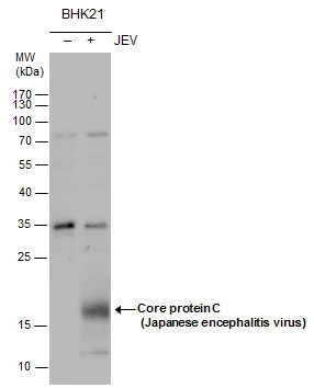
Core protein C (JEV) antibody detects Core protein C (JEV) protein by western blot analysis. Un-infected (-) and infected (+, JEV infection) BHK-21 whole cell extracts (30 μg) were separated by 15% SDS-PAGE, and the membrane was blotted with Core protein C (JEV) antibody (GTX131368) at a dilution of 1:3000 and developed with Trident femto Western HRP Substrate (GTX14698). The HRP-conjugated anti-rabbit IgG antibody (GTX213110-01) was used to detect the primary antibody.
 antibody detects Core protein C (Japanese encephalitis virus) by immunofluorescent analysis. Samples: BHK-21 cells mock (left) and infected with Japanese encephalitis virus (right) were fixed in methanol. Green: Core protein C (Japanese encephalitis virus) stained by core protein C (Japanese encephalitis virus) antibody (GTX131368) diluted at 1:3000. Blue: Hoechst 33342 staining.")
Core protein C (JEV) antibody detects Core protein C (JEV) protein by western blot analysis. Un-infected (-) and infected (+, JEV infection) BHK-21 whole cell extracts (30 μg) were separated by 15% SDS-PAGE, and the membrane was blotted with Core protein C (JEV) antibody (GTX131368) at a dilution of 1:3000 and developed with Trident femto Western HRP Substrate (GTX14698). The HRP-conjugated anti-rabbit IgG antibody (GTX213110-01) was used to detect the primary antibody.
Japanese encephalitis virus Core protein C antibody
GTX131368
ApplicationsImmunoFluorescence, Western Blot, ImmunoCytoChemistry
Product group Antibodies
ReactivityVirus
Overview
- SupplierGeneTex
- Product NameJapanese encephalitis virus Core protein C antibody
- Delivery Days Customer9
- Application Supplier NoteWB: 1:500-1:3000. ICC/IF: 1:100-1:3000. *Optimal dilutions/concentrations should be determined by the researcher.Not tested in other applications.
- ApplicationsImmunoFluorescence, Western Blot, ImmunoCytoChemistry
- CertificationResearch Use Only
- ClonalityPolyclonal
- Concentration1 mg/ml
- ConjugateUnconjugated
- HostRabbit
- IsotypeIgG
- Scientific DescriptionThe small proteins NS2A, NS4A and NS4B are hydrophobic, suggesting a possible membrane-related function. Non-structural protein 2B is a required cofactor for the serine protease function of NS3. Serine protease NS3 displays three enzymatic activities: serine protease, NTPase and RNA helicase. NS3 serine protease, in association with NS2B, cleaves the polyprotein. RNA-directed RNA polymerase NS5 replicates the viral (+) and (-) genome, and performs the capping of genomes in the cytoplasm. NS5 methylates viral RNA cap at guanine N-7 and ribose 2-O positions.
- ReactivityVirus
- Storage Instruction-20°C or -80°C,2°C to 8°C
- UNSPSC12352203
References
- Ma X, Li C, Xia Q, et al. Construction of a Recombinant Japanese Encephalitis Virus with a Hemagglutinin-Tagged NS2A: A Model for an Analysis of Biological Characteristics and Functions of NS2A during Viral Infection. Viruses. 2022,14(4). doi: 10.3390/v14040706Read this paper
- Sehrawat S, Khasa R, Deb A, et al. Valosin-containing protein/p97 plays critical roles in the Japanese encephalitis virus life cycle. J Virol. 2021,95(11):pii: JVI.02336-20. doi: 10.1128/JVI.02336-20.Read this paper
- Sarkar R, Sharma KB, Kumari A, et al. Japanese encephalitis virus capsid protein interacts with non-lipidated MAP1LC3 on replication membranes and lipid droplets. J Gen Virol. 2021,102(1). doi: 10.1099/jgv.0.001508Read this paper