

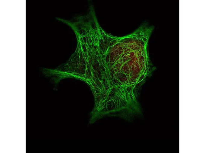
Immunofluorescence Microscopy of GeneTex Anti-Keratin antibody (GTX48825) was used with GeneTex Dylight 488 goat anti-mouse (shown in green) to detect Keratin by Immunofluorescence. In the same experiment, GeneTex polyclonal Anti-HDAC-1 antibody was used with Anti-Rabbit IgG (shown in red) to detect HDAC-1.
![Western blot using GeneTex Immunochemical's Mouse Mab-anti-Keratin antibody. This antibody recognizes a single 56 kDa band corresponding to human keratin as confirmed by the position of molecular weight markers (not shown). Approximatley 100 μg of keratin from human epidermis was applied under reducing conditions. 1:400 dilution of Mab anti-Keratin was used for 2h followed by detection using a 1:5,000 dilution of IRDyeTM800 conjugated Goat anti-Mouse IgG [H&L] and visualization using the OdysseyR Infrared Imaging System developed by LI-COR.](https://www.genetex.com/upload/website/prouct_img/normal/GTX48825/GTX48825_20160330_WB_w_23060823_385.webp "Western blot using GeneTex Immunochemical's Mouse Mab-anti-Keratin antibody. This antibody recognizes a single 56 kDa band corresponding to human keratin as confirmed by the position of molecular weight markers (not shown). Approximatley 100 μg of keratin from human epidermis was applied under reducing conditions. 1:400 dilution of Mab anti-Keratin was used for 2h followed by detection using a 1:5,000 dilution of IRDyeTM800 conjugated Goat anti-Mouse IgG [H&L] and visualization using the OdysseyR Infrared Imaging System developed by LI-COR.")
![Immunofluorescence microscopy using GeneTex Immunochemical's Mouse Mab-anti-Keratin antibody. Confocal slices of HeLa cells are between 0.5 and 0.6 um where the image is taken near the bottom of the cell. Use FITC a 1:2,000 dilution of FITC-conjugated Goat anti-Mouse IgG [H&L] for detection.](https://www.genetex.com/upload/website/prouct_img/normal/GTX48825/GTX48825_20160330_ICCIF_1_w_23060823_288.webp "Immunofluorescence microscopy using GeneTex Immunochemical's Mouse Mab-anti-Keratin antibody. Confocal slices of HeLa cells are between 0.5 and 0.6 um where the image is taken near the bottom of the cell. Use FITC a 1:2,000 dilution of FITC-conjugated Goat anti-Mouse IgG [H&L] for detection.")
![ICC/IF analysis of HeLa cells using GTX48825 pan Cytokeratin antibody [C-11].](https://www.genetex.com/upload/website/prouct_img/normal/GTX48825/GTX48825_20240423_ICCIF_323_24042320_116.webp "ICC/IF analysis of HeLa cells using GTX48825 pan Cytokeratin antibody [C-11].")
![WB analysis of recombinant human pan Cytokeratin protein using GTX48825 pan Cytokeratin antibody [C-11]. Loading : 100 ng Dilution : 1:400](https://www.genetex.com/upload/website/prouct_img/normal/GTX48825/GTX48825_20240423_WB_324_24042320_534.webp "WB analysis of recombinant human pan Cytokeratin protein using GTX48825 pan Cytokeratin antibody [C-11]. Loading : 100 ng Dilution : 1:400")
Immunofluorescence Microscopy of GeneTex Anti-Keratin antibody (GTX48825) was used with GeneTex Dylight 488 goat anti-mouse (shown in green) to detect Keratin by Immunofluorescence. In the same experiment, GeneTex polyclonal Anti-HDAC-1 antibody was used with Anti-Rabbit IgG (shown in red) to detect HDAC-1.
pan Cytokeratin antibody [C-11]
GTX48825
ApplicationsImmunoFluorescence, ImmunoPrecipitation, Western Blot, ELISA, ImmunoCytoChemistry, ImmunoHistoChemistry, ImmunoHistoChemistry Paraffin, Other Application
Product group Antibodies
ReactivityHuman
TargetKRT6C
Overview
- SupplierGeneTex
- Product Namepan Cytokeratin antibody [C-11]
- Delivery Days Customer9
- Application Supplier NoteWB: 1:50-1:200. ICC/IF: 1:50-1:200. IHC-P: 1:50-1:200. IP: 1:100. ELISA: 1:5000-1:20000. *Optimal dilutions/concentrations should be determined by the researcher.Not tested in other applications.
- ApplicationsImmunoFluorescence, ImmunoPrecipitation, Western Blot, ELISA, ImmunoCytoChemistry, ImmunoHistoChemistry, ImmunoHistoChemistry Paraffin, Other Application
- CertificationResearch Use Only
- ClonalityMonoclonal
- Clone IDC-11
- Concentration1.3 mg/ml
- ConjugateUnconjugated
- Gene ID286887
- Target nameKRT6C
- Target descriptionkeratin 6C
- Target synonymsK6E, KRT6E, PPKNEFD, keratin, type II cytoskeletal 6C, CK-6C, CK-6E, K6C, cytokeratin-6C, cytokeratin-6E, keratin 6C, type II, keratin 6E, keratin K6h, type-II keratin Kb12
- HostMouse
- IsotypeIgG1
- Protein IDP48668
- Protein NameKeratin, type II cytoskeletal 6C
- Scientific DescriptionCytokeratins (CK) are intermediate filaments of epithelial cells, both in keratinizing tissue (i.e. skin) and non-keratinizing cells (i.e. mesothelial)
- ReactivityHuman
- Storage Instruction-20°C or -80°C,2°C to 8°C
- UNSPSC41116161
References
- Extracellular vesicle analysis of plasma allows differential diagnosis of atypical pancreatic serous cystadenoma.Read this paper
- Broad-spectrum immunohistochemical epithelial markers: a review. Ordonez NG, 2013 Jul, Hum PatholRead this paper
Datasheet
Related products
Product group Antibodies
KRT6C AntibodyCSB-PA216033
ApplicationsELISA, ImmunoHistoChemistry
ReactivityHuman, Mouse, Rat
TargetKRT6C
- SizePrice
Product group Antibodies
References
ApplicationsWestern Blot, ELISA
ReactivityHuman
TargetKRT6C
- SizePrice
Product group Antibodies

ApplicationsWestern Blot
ReactivityHuman
TargetKRT6C
- SizePrice
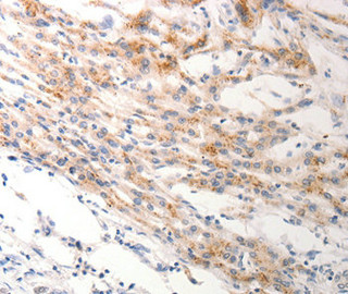
![IHC-P analysis of guinea pig breast carcinoma tissue using GTX27753 pan Cytokeratin antibody [C-11].](https://www.genetex.com/upload/website/prouct_img/normal/GTX27753/GTX27753_20191028_IHC-P_1_w_23060722_247.webp)
Product group Antibodies
References
pan Cytokeratin antibody [C-11]GTX27753
ApplicationsFlow Cytometry, ImmunoFluorescence, ImmunoPrecipitation, Western Blot, ImmunoCytoChemistry, ImmunoHistoChemistry, ImmunoHistoChemistry Paraffin
ReactivityGuinea Pig, Human, Mouse, Rat
- SizePrice
Product group Antibodies
ApplicationsWestern Blot, ImmunoHistoChemistry
TargetKRT6C
- SizePrice
Product group Antibodies
Keratin 6 Recombinant AntibodyBSM-60235R
ApplicationsImmunoFluorescence, Western Blot, ImmunoCytoChemistry, ImmunoHistoChemistry, ImmunoHistoChemistry Frozen, ImmunoHistoChemistry Paraffin
ReactivityHuman, Mouse, Rat
TargetKRT6C
- SizePrice