Bio-Connect

KRT8 Antibody (OAAF01014)

OAAF01014
Aviva Systems Biology
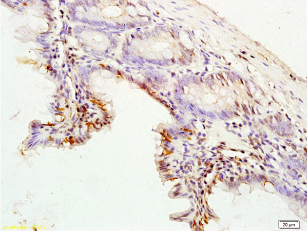
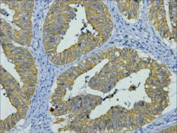
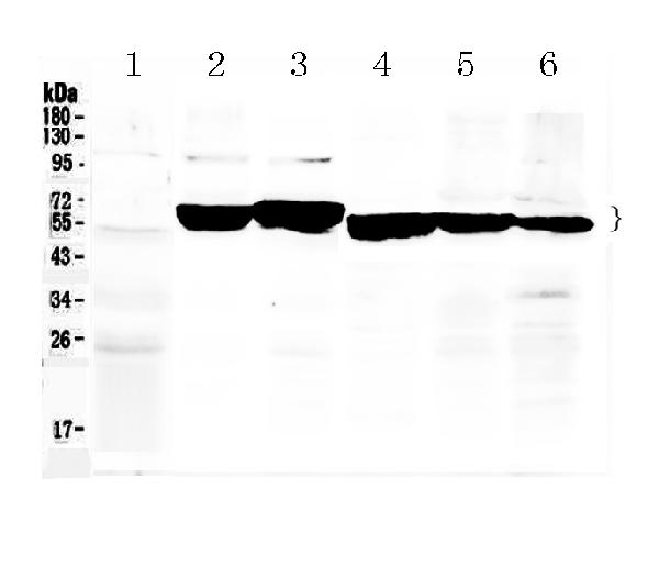
ApplicationsImmunoFluorescence, Western Blot, ImmunoHistoChemistry
Product group Antibodies
TargetKRT8
Sign in to order and to see your custom pricing.
Large volume orders?
Order with a bulk request

Overview

  • Supplier
    Aviva Systems Biology
  • Product Name
    KRT8 Antibody (OAAF01014)
  • Delivery Days Customer
    23
  • Applications
    ImmunoFluorescence, Western Blot, ImmunoHistoChemistry
  • Certification
    Research Use Only
  • Clonality
    Polyclonal
  • Concentration
    1 mg/ml
  • Format
    Liquid. PBS (without Mg2+ and Ca2+), pH 7.4, 150mM NaCl, 0.02% sodium azide and 50% glycerol.
  • Gene ID3856
  • Target name
    KRT8
  • Target description
    keratin 8
  • Target synonyms
    CARD2; CK8; CK-8; CYK8; cytokeratin-8; K2C8; K8; keratin 8, type II; keratin, type II cytoskeletal 8; KO; type-II keratin Kb8
  • Host
    Rabbit
  • Isotype
    IgG
  • Storage Instruction
    -20°C
  • UNSPSC
    12352203

Related products

Formalin-fixed and rat colon tissue labeled with Anti-CK8 Polyclonal Antibody, Unconjugated (bs-1106R) at 1:200 followed by conjugation to the secondary antibody and DAB staining
Product group Antibodies
References
Bioss Antibodies
ApplicationsFlow Cytometry, ImmunoFluorescence, Western Blot, ELISA, ImmunoCytoChemistry, ImmunoHistoChemistry, ImmunoHistoChemistry Frozen, ImmunoHistoChemistry Paraffin
TargetKRT8
  • SizePrice
Product group Antibodies
Biomatik
ApplicationsImmunoFluorescence, Western Blot, ELISA, ImmunoHistoChemistry
TargetKRT8
  • SizePrice
Product group Antibodies
Assay Genie
TargetKRT8
  • SizePrice
Product group Antibodies
LifeSpan BioSciences
ApplicationsWestern Blot, ELISA
TargetKRT8
  • SizePrice
Product group Antibodies
Biosynth
  • SizePrice
Product group Antibodies
RayBiotech
ApplicationsWestern Blot, ELISA
  • SizePrice
IHC staining of human colon cancer tissue, diluted at 1:200.
Product group Antibodies
Cusabio
ApplicationsWestern Blot, ELISA, ImmunoHistoChemistry
ReactivityHuman, Mouse, Rat
TargetKRT8
  • SizePrice
Immunohistochemical staining of human small intestine shows strong membranous positivity in glandular cells.
Product group Antibodies
Atlas Antibodies
ApplicationsWestern Blot, ImmunoCytoChemistry, ImmunoHistoChemistry
ReactivityHuman
TargetKRT8
  • SizePrice
Product group Antibodies
Nordic-MUbio
ApplicationsWestern Blot, ImmunoHistoChemistry, ImmunoHistoChemistry Frozen
TargetKRT8
  • SizePrice
Figure 1. Western blot analysis of Cytokeratin 8 using anti-Cytokeratin 8 antibody (A01421). Electrophoresis was performed on a 5-20% SDS-PAGE gel at 70V (Stacking gel) / 90V (Resolving gel) for 2-3 hours. The sample well of each lane was loaded with 50ug of sample under reducing conditions. Lane 1: rat liver tissue lysates, Lane 2: mouse ovary tissue lysates, Lane 3: mouse liver tissue lysates, Lane 4: MCF-7 whole cell lysates, Lane 5: A549 whole cell lysates, Lane 6: HELA whole Cell lysates. After Electrophoresis, proteins were transferred to a Nitrocellulose membrane at 150mA for 50-90 minutes. Blocked the membrane with 5% Non-fat Milk/ TBS for 1.5 hour at RT. The membrane was incubated with rabbit anti-Cytokeratin 8 antigen affinity purified polyclonal antibody (Catalog # A01421) at 0.5 microg/mL overnight at 4°C, then washed with TBS-0.1%Tween 3 times with 5 minutes each and probed with a goat anti-rabbit IgG-HRP secondary antibody at a dilution of 1:10000 for 1.5 hour at RT. The signal is developed using an Enhanced Chemiluminescent detection (ECL) kit (Catalog # EK1002) with Tanon 5200 system. A specific band was detected for Cytokeratin 8 at approximately 54KD, 56KD. The expected band size for Cytokeratin 8 is at 54KD.
Product group Antibodies
Boster Bio
ApplicationsWestern Blot, ImmunoHistoChemistry
TargetKRT8
  • SizePrice