

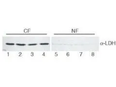
Western blot analysis shows GeneTex's Anti-Lactate Dehydrogenase antibody detects LDH in HeLa cell extracts. Reactivity with LDH is observed in the cytoplasmic fraction (CF) and little to no reactivity in the nuclear fraction (NF). 30 μg was loaded per lane. Comparison to a molecular weight marker (not shown) indicates a single band of ~36 kDa corresponding to the expected molecular weight for the protein. The blot was incubated with 1:400 for overnight at 4oC. Secondary antibody at 1:10,000 for 45 min at RT.


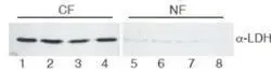
Western blot analysis shows GeneTex's Anti-Lactate Dehydrogenase antibody detects LDH in HeLa cell extracts. Reactivity with LDH is observed in the cytoplasmic fraction (CF) and little to no reactivity in the nuclear fraction (NF). 30 μg was loaded per lane. Comparison to a molecular weight marker (not shown) indicates a single band of ~36 kDa corresponding to the expected molecular weight for the protein. The blot was incubated with 1:400 for overnight at 4oC. Secondary antibody at 1:10,000 for 45 min at RT.
Lactate Dehydrogenase antibody
GTX48868
ApplicationsImmunoFluorescence, ImmunoPrecipitation, Western Blot, ELISA, ImmunoCytoChemistry
Product group Antibodies
ReactivityHuman, Rabbit
Overview
- SupplierGeneTex
- Product NameLactate Dehydrogenase antibody
- Delivery Days Customer9
- Application Supplier NoteWB: 1:500-1:5000. IP: 1:100. ELISA: 1:5000-1:20000. *Optimal dilutions/concentrations should be determined by the researcher.Not tested in other applications.
- ApplicationsImmunoFluorescence, ImmunoPrecipitation, Western Blot, ELISA, ImmunoCytoChemistry
- CertificationResearch Use Only
- ClonalityPolyclonal
- Concentration82 mg/ml
- ConjugateUnconjugated
- HostGoat
- IsotypeIgG
- Scientific DescriptionLactate dehydrogenase is also known as L-lactate dehydrogenase A chain, LDH-A, LDH muscle subunit and LDH-M. Two isozymes of LDH occur in mammals, LDH-M and LDH-H which come together to form a homotetramer of 36 kDa subunits. Every LDH molecule consists of four subunits, where each subunit is either H each M (based on their electrophoretic properties.) There are, therefore, five LDH isotypes: LDH-1 (4H) - in the heart, LDH-2 (3H1M) - in the reticuloendothelial system, LDH-3 (2H2M) - in the lungs, LDH-4 (1H3M) - in the kidneys and LDH-5 (4M) - in the liver and striated muscle. Usually LDH-2 is the predominant form in the serum. An LDH-1 level higher than the LDH-2 level (a flipped pattern) suggests myocardial infarction (damage to heart tissues releases heart LDH, which is rich in LDH-1, into the bloodstream). In general, LDH is often used as a marker of tissue breakdown. LDH shows a cytoplasmic localization.
- ReactivityHuman, Rabbit
- Storage Instruction-20°C or -80°C,2°C to 8°C
- UNSPSC41116161
Datasheet
Related products

Product group Antibodies
Lactate Dehydrogenase antibodyGTX22101
ApplicationsImmunoFluorescence, ImmunoPrecipitation, Western Blot, ELISA, ImmunoCytoChemistry
ReactivityHuman, Rabbit
TargetLDHA
- SizePrice