

ICC/IF analysis of PFA-fixed HeLa cells with/without Chloroquine (50 μM, 37oC, 20 hrs) treatment using GTX00949 LC3B antibody [GT1187]. Right : untreated HeLa cells Left : HeLa cells with 50 μM Chloroquine (20 hrs, 37oC) treatment Orange : Primary antibody Blue : DAPI Dilution : 1:100
![ICC/IF analysis of PFA-fixed NIH-3T3 cells with/without Chloroquine (50 μM, 37oC, 20 hrs) treatment using GTX00949 LC3B antibody [GT1187]. Right : untreated NIH-3T3 cells Left : NIH-3T3 cells with 50 μM Chloroquine (20 hrs, 37oC) treatment Orange : Primary antibody Blue : DAPI Dilution : 1:100](https://www.genetex.com/upload/website/prouct_img/normal/GTX00949/GTX00949_20200327_ICC-IF_44_w_23053121_674.webp "ICC/IF analysis of PFA-fixed NIH-3T3 cells with/without Chloroquine (50 μM, 37oC, 20 hrs) treatment using GTX00949 LC3B antibody [GT1187]. Right : untreated NIH-3T3 cells Left : NIH-3T3 cells with 50 μM Chloroquine (20 hrs, 37oC) treatment Orange : Primary antibody Blue : DAPI Dilution : 1:100")
![IHC-P analysis of Mouse spinal cord tissue section using GTX00949 LC3B antibody [GT1187]. Dilution : 1:100](https://www.genetex.com/upload/website/prouct_img/normal/GTX00949/GTX00949_20200327_IHC-P_41_w_23053121_323.webp "IHC-P analysis of Mouse spinal cord tissue section using GTX00949 LC3B antibody [GT1187]. Dilution : 1:100")
![IHC-P analysis of Rat brain tissue section using GTX00949 LC3B antibody [GT1187]. Dilution : 1:100](https://www.genetex.com/upload/website/prouct_img/normal/GTX00949/GTX00949_20200327_IHC-P_39_w_23053121_454.webp "IHC-P analysis of Rat brain tissue section using GTX00949 LC3B antibody [GT1187]. Dilution : 1:100")
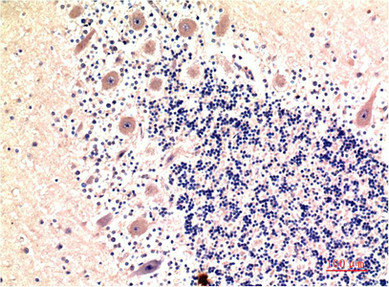
![IHC-P analysis of Human brain tissue section using GTX00949 LC3B antibody [GT1187]. Dilution : 1:100](https://www.genetex.com/upload/website/prouct_img/normal/GTX00949/GTX00949_20200327_IHC-P_40_w_23053121_926.webp "IHC-P analysis of Human brain tissue section using GTX00949 LC3B antibody [GT1187]. Dilution : 1:100")
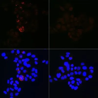
![ICC/IF analysis of PFA-fixed C6 cells with/without Chloroquine (50 μM, 37oC, 20 hrs) treatment using GTX00949 LC3B antibody [GT1187]. Right : untreated C6 cells Left : C6 cells with 50 μM Chloroquine (20 hrs, 37oC) treatment Orange : Primary antibody Blue : DAPI Dilution : 1:100](https://www.genetex.com/upload/website/prouct_img/normal/GTX00949/GTX00949_20200327_ICC-IF_42_w_23053121_487.webp "ICC/IF analysis of PFA-fixed C6 cells with/without Chloroquine (50 μM, 37oC, 20 hrs) treatment using GTX00949 LC3B antibody [GT1187]. Right : untreated C6 cells Left : C6 cells with 50 μM Chloroquine (20 hrs, 37oC) treatment Orange : Primary antibody Blue : DAPI Dilution : 1:100")
![Untreated (–) and treated (+) HepG2 whole cell extracts (30 μg) were separated by 15% SDS-PAGE, and the membrane was blotted with LC3B antibody [GT1187] (GTX00949) diluted at 1:500. The HRP-conjugated anti-rabbit IgG antibody (GTX213110-01) was used to detect the primary antibody.](https://www.genetex.com/upload/website/prouct_img/normal/GTX00949/GTX00949_40000000144_20200306_WB_treatment_Thapsigargin_w_23053121_647.webp "Untreated (–) and treated (+) HepG2 whole cell extracts (30 μg) were separated by 15% SDS-PAGE, and the membrane was blotted with LC3B antibody [GT1187] (GTX00949) diluted at 1:500. The HRP-conjugated anti-rabbit IgG antibody (GTX213110-01) was used to detect the primary antibody.")
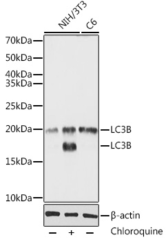
![WB analysis of C6 and NIH-3T3 cell extracts with/without Chloroquine (50 μM, 37oC, 20 hrs) treatment using GTX00949 LC3B antibody [GT1187]. Dilution : 1:1000 Loading : 25μg](https://www.genetex.com/upload/website/prouct_img/normal/GTX00949/GTX00949_20200327_WB_2_w_23053121_126.webp "WB analysis of C6 and NIH-3T3 cell extracts with/without Chloroquine (50 μM, 37oC, 20 hrs) treatment using GTX00949 LC3B antibody [GT1187]. Dilution : 1:1000 Loading : 25μg")
![WB analysis of wild-type (WT) and LC3B knockout (KO) 293T cell extracts with/without Chloroquine (50 μM, 37oC, 20 hrs) treatment using GTX00949 LC3B antibody [GT1187]. Dilution : 1:1000 Loading : 25μg](https://www.genetex.com/upload/website/prouct_img/normal/GTX00949/GTX00949_20200327_WB_1_w_23053121_206.webp "WB analysis of wild-type (WT) and LC3B knockout (KO) 293T cell extracts with/without Chloroquine (50 μM, 37oC, 20 hrs) treatment using GTX00949 LC3B antibody [GT1187]. Dilution : 1:1000 Loading : 25μg")
ICC/IF analysis of PFA-fixed HeLa cells with/without Chloroquine (50 μM, 37oC, 20 hrs) treatment using GTX00949 LC3B antibody [GT1187]. Right : untreated HeLa cells Left : HeLa cells with 50 μM Chloroquine (20 hrs, 37oC) treatment Orange : Primary antibody Blue : DAPI Dilution : 1:100
LC3B antibody [GT1187]
GTX00949
ApplicationsImmunoFluorescence, Western Blot, ImmunoCytoChemistry, ImmunoHistoChemistry, ImmunoHistoChemistry Paraffin
Product group Antibodies
ReactivityHuman, Mouse, Rat
TargetMAP1LC3B
Overview
- SupplierGeneTex
- Product NameLC3B antibody [GT1187]
- Delivery Days Customer9
- Application Supplier NoteWB: 1:500 - 1:2000. ICC/IF: 1:50 - 1:200. IHC-P: 1:50 - 1:200. *Optimal dilutions/concentrations should be determined by the researcher.Not tested in other applications.
- ApplicationsImmunoFluorescence, Western Blot, ImmunoCytoChemistry, ImmunoHistoChemistry, ImmunoHistoChemistry Paraffin
- CertificationResearch Use Only
- ClonalityMonoclonal
- Clone IDGT1187
- ConjugateUnconjugated
- Gene ID81631
- Target nameMAP1LC3B
- Target descriptionmicrotubule associated protein 1 light chain 3 beta
- Target synonymsATG8F, LC3B, MAP1A/1BLC3, MAP1LC3B-a, microtubule-associated protein 1 light chain 3 beta, MAP1 light chain 3-like protein 2, MAP1A/MAP1B LC3 B, MAP1A/MAP1B light chain 3 B, autophagy-related ubiquitin-like modifier LC3 B, microtubule-associated proteins 1A/1B light chain 3B
- HostRabbit
- IsotypeIgG
- Protein IDQ9GZQ8
- Protein NameMicrotubule-associated protein 1 light chain 3 beta
- Scientific DescriptionThe product of this gene is a subunit of neuronal microtubule-associated MAP1A and MAP1B proteins, which are involved in microtubule assembly and important for neurogenesis. Studies on the rat homolog implicate a role for this gene in autophagy, a process that involves the bulk degradation of cytoplasmic component. [provided by RefSeq, Jul 2008]
- ReactivityHuman, Mouse, Rat
- Storage Instruction-20°C or -80°C,2°C to 8°C
- UNSPSC41116161
Datasheet
Related products
Product group Antibodies
Anti-LC3B AntibodyA80596
ApplicationsImmunoFluorescence, Western Blot, ImmunoCytoChemistry, ImmunoHistoChemistry
ReactivityHuman, Mouse, Rat
- SizePrice
Product group Antibodies
Anti-MAP1LC3B Antibody144-11282
ApplicationsImmunoFluorescence, Western Blot, ImmunoHistoChemistry
ReactivityHuman, Mouse, Porcine, Rat
TargetMAP1LC3B
- SizePrice
Product group Antibodies

Cleaved LC3B AntibodyABX029981
ApplicationsImmunoFluorescence, ELISA, ImmunoCytoChemistry
- SizePrice
Product group Antibodies

MAP1LC3B / LC3B Antibody (clone 6E4)LS-C768006
ApplicationsImmunoHistoChemistry, ImmunoHistoChemistry Paraffin
ReactivityHuman
TargetMAP1LC3B
- SizePrice
Product group Antibodies
MAP1LC3B Monoclonal AntibodyCSB-MA171423
ApplicationsELISA, ImmunoHistoChemistry
ReactivityHuman
TargetMAP1LC3B
- SizePrice
Product group Antibodies
Map1Lc3B Polyclonal AntibodyCAC09906
ApplicationsELISA, ImmunoHistoChemistry
TargetMAP1LC3B
- SizePrice
Product group Antibodies
References
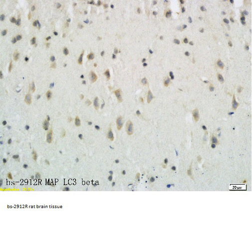
LC3B Polyclonal AntibodyBS-2912R
ApplicationsImmunoFluorescence, ImmunoHistoChemistry, ImmunoHistoChemistry Frozen, ImmunoHistoChemistry Paraffin
ReactivityBovine, Canine, Chicken, Equine, Human, Mouse, Porcine, Rabbit, Rat, Zebra Fish
TargetMAP1LC3B
- SizePrice

Product group Antibodies
References
ApplicationsImmunoFluorescence, ImmunoPrecipitation, Western Blot, ImmunoCytoChemistry
ReactivityHuman, Mouse, Rat
TargetMAP1LC3B
- SizePrice

Product group Antibodies
LC3B antibodyGTX31899
ApplicationsWestern Blot, ELISA, ImmunoHistoChemistry, ImmunoHistoChemistry Paraffin
ReactivityHuman, Mouse, Rat
TargetMAP1LC3B
- SizePrice