

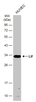
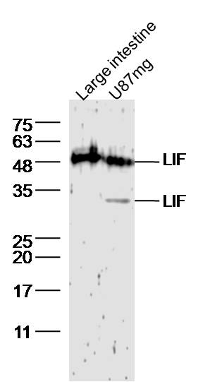
Whole cell extract (30 μg) was separated by 12% SDS-PAGE, and the membrane was blotted with LIF antibody (GTX101021) diluted at 1:1000. The HRP-conjugated anti-rabbit IgG antibody (GTX213110-01) was used to detect the primary antibody.
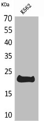
![Whole cell extract (30 μg) was separated by 12% SDS-PAGE, and the membranes were blotted with LIF antibody [N1C3] (GTX101021) diluted at 1:500 and competitor's antibody (sc-1336) diluted at 1:500. The HRP-conjugated anti-rabbit IgG antibody (GTX213110-01) was used to detect the primary antibody.](https://www.genetex.com/upload/website/prouct_img/normal/GTX101021/GTX101021_42872_20171006_WB_competitor_watermark_w_23060100_786.webp "Whole cell extract (30 μg) was separated by 12% SDS-PAGE, and the membranes were blotted with LIF antibody [N1C3] (GTX101021) diluted at 1:500 and competitor's antibody (sc-1336) diluted at 1:500. The HRP-conjugated anti-rabbit IgG antibody (GTX213110-01) was used to detect the primary antibody.")
 were separated by 12% SDS-PAGE, and the membrane was blotted with LIF antibody (GTX101021) diluted at 1:1000. The HRP-conjugated anti-rabbit IgG antibody (GTX213110-01) was used to detect the primary antibody.")
Whole cell extract (30 μg) was separated by 12% SDS-PAGE, and the membrane was blotted with LIF antibody (GTX101021) diluted at 1:1000. The HRP-conjugated anti-rabbit IgG antibody (GTX213110-01) was used to detect the primary antibody.
LIF antibody
GTX101021
ApplicationsWestern Blot
Product group Antibodies
ReactivityHuman
TargetLIF
Overview
- SupplierGeneTex
- Product NameLIF antibody
- Delivery Days Customer9
- Application Supplier NoteWB: 1:500-1:3000. *Optimal dilutions/concentrations should be determined by the researcher.Not tested in other applications.
- ApplicationsWestern Blot
- CertificationResearch Use Only
- ClonalityPolyclonal
- Concentration1.07 mg/ml
- ConjugateUnconjugated
- Gene ID3976
- Target nameLIF
- Target descriptionLIF interleukin 6 family cytokine
- Target synonymsCDF, DIA, HILDA, MLPLI, leukemia inhibitory factor, D factor, cholinergic differentiation factor, differentiation inhibitory activity, differentiation-inducing factor, differentiation-stimulating factor, hepatocyte-stimulating factor III, human interleukin in DA cells, melanoma-derived LPL inhibitor
- HostRabbit
- IsotypeIgG
- Protein IDP15018
- Protein NameLeukemia inhibitory factor
- Scientific DescriptionThe protein encoded by this gene is a pleiotropic cytokine with roles in several different systems. It is involved in the induction of hematopoietic differentiation in normal and myeloid leukemia cells, induction of neuronal cell differentiation, regulator of mesenchymal to epithelial conversion during kidney development, and may also have a role in immune tolerance at the maternal-fetal interface. [provided by RefSeq]
- ReactivityHuman
- Storage Instruction-20°C or -80°C,2°C to 8°C
- UNSPSC41116161
Datasheet
Related products
Product group Antibodies
ApplicationsImmunoPrecipitation, Western Blot, ImmunoCytoChemistry, ImmunoHistoChemistry
ReactivityRat
TargetLIF
- SizePrice
Product group Antibodies
LIF AntibodyCSB-PA004761
ApplicationsWestern Blot, ELISA
ReactivityHuman
TargetLIF
- SizePrice
Product group Antibodies
Anti-LIF AntibodyA101615
ApplicationsWestern Blot, ELISA
ReactivityHuman
- SizePrice
Product group Antibodies
Anti-LIF Antibody144-62539
ApplicationsImmunoHistoChemistry
ReactivityHuman
TargetLIF
- SizePrice
Product group Antibodies

LIF Antibody (Preservative Free)LS-C820626
ApplicationsELISA
ReactivityMouse
TargetLIF
- SizePrice
Product group Antibodies

Goat anti-LIFEB11863
ApplicationsFlow Cytometry, ELISA, ImmunoHistoChemistry
ReactivityHuman
TargetLIF
- SizePrice

Product group Antibodies
Anti-LIF AntibodyHPA018844
ApplicationsWestern Blot, ImmunoCytoChemistry, ImmunoHistoChemistry
ReactivityHuman
TargetLIF
- SizePrice
Product group Antibodies
Anti-LIF Antibody Picoband(r)PB9036-CARRIER-FREE
ApplicationsWestern Blot, ImmunoHistoChemistry
ReactivityHuman
TargetLIF
- SizePrice
Product group Antibodies
LIF antibody [6H31]GTX52909
ApplicationsImmunoHistoChemistry, ImmunoHistoChemistry Paraffin, Neutralisation/Blocking
ReactivityHuman
TargetLIF
- SizePrice
Product group Antibodies
LIF Polyclonal AntibodyBS-1058R
ApplicationsImmunoFluorescence, Western Blot, ELISA, ImmunoCytoChemistry, ImmunoHistoChemistry, ImmunoHistoChemistry Frozen, ImmunoHistoChemistry Paraffin
TargetLIF
- SizePrice