

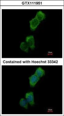
Immunofluorescence analysis of methanol-fixed A431, using LOK(GTX111951) antibody at 1:500 dilution.
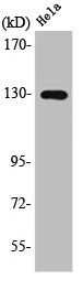
![Wild-type (WT) and LOK knockout (KO) HeLa cell extracts (30 μg) were separated by 5% SDS-PAGE, and the membrane was blotted with LOK antibody [N1N2], N-term (GTX111951) diluted at 1:2000. The HRP-conjugated anti-rabbit IgG antibody (GTX213110-01) was used to detect the primary antibody.](https://www.genetex.com/upload/website/prouct_img/normal/GTX111951/GTX111951_40464_20170601_WB_KO_watermark_w_23060500_910.webp "Wild-type (WT) and LOK knockout (KO) HeLa cell extracts (30 μg) were separated by 5% SDS-PAGE, and the membrane was blotted with LOK antibody [N1N2], N-term (GTX111951) diluted at 1:2000. The HRP-conjugated anti-rabbit IgG antibody (GTX213110-01) was used to detect the primary antibody.")
 antibody at 1:250 dilution.
Antigen Retrieval: Trilogy? (EDTA based, pH 8.0) buffer, 15min")
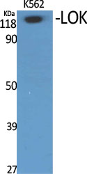
![Various whole cell extracts (30 μg) were separated by 5% SDS-PAGE, and the membrane was blotted with LOK antibody [N1N2], N-term (GTX111951) diluted at 1:2000. The HRP-conjugated anti-rabbit IgG antibody (GTX213110-01) was used to detect the primary antibody, and the signal was developed with Trident ECL plus-Enhanced.](https://www.genetex.com/upload/website/prouct_img/normal/GTX111951/GTX111951_40464_20231027_WB_23103019_668.webp "Various whole cell extracts (30 μg) were separated by 5% SDS-PAGE, and the membrane was blotted with LOK antibody [N1N2], N-term (GTX111951) diluted at 1:2000. The HRP-conjugated anti-rabbit IgG antibody (GTX213110-01) was used to detect the primary antibody, and the signal was developed with Trident ECL plus-Enhanced.")
Immunofluorescence analysis of methanol-fixed A431, using LOK(GTX111951) antibody at 1:500 dilution.
LOK antibody [N1N2], N-term
GTX111951
ApplicationsImmunoFluorescence, Western Blot, ImmunoCytoChemistry, ImmunoHistoChemistry, ImmunoHistoChemistry Paraffin
Product group Antibodies
ReactivityHuman
TargetSTK10
Overview
- SupplierGeneTex
- Product NameLOK antibody [N1N2], N-term
- Delivery Days Customer9
- Application Supplier NoteWB: 1:500-1:3000. ICC/IF: 1:100-1:1000. IHC-P: 1:100-1:1000. *Optimal dilutions/concentrations should be determined by the researcher.Not tested in other applications.
- ApplicationsImmunoFluorescence, Western Blot, ImmunoCytoChemistry, ImmunoHistoChemistry, ImmunoHistoChemistry Paraffin
- CertificationResearch Use Only
- ClonalityPolyclonal
- Concentration1 mg/ml
- ConjugateUnconjugated
- Gene ID6793
- Target nameSTK10
- Target descriptionserine/threonine kinase 10
- Target synonymsLOK, PRO2729, serine/threonine-protein kinase 10, lymphocyte-oriented kinase
- HostRabbit
- IsotypeIgG
- Protein IDO94804
- Protein NameSerine/threonine-protein kinase 10
- Scientific DescriptionThis gene encodes a member of the Ste20 family of serine/threonine protein kinases, and is similar to several known polo-like kinase kinases. The protein can associate with and phosphorylate polo-like kinase 1, and overexpression of a kinase-dead version of the protein interferes with normal cell cycle progression. The kinase can also negatively regulate interleukin 2 expression in T-cells via the mitogen activated protein kinase kinase 1 pathway. [provided by RefSeq]
- ReactivityHuman
- Storage Instruction-20°C or -80°C,2°C to 8°C
- UNSPSC12352203
Datasheet
Related products
Product group Antibodies

STK10 / LOK AntibodyLS-C749262
ApplicationsWestern Blot
ReactivityHuman
TargetSTK10
- SizePrice
Product group Antibodies
Anti-STK10 AntibodyA100760
ApplicationsWestern Blot, ELISA
ReactivityHuman
- SizePrice
Product group Antibodies
STK10 AntibodyCSB-PA003160
ApplicationsWestern Blot, ELISA
ReactivityHuman
TargetSTK10
- SizePrice

Product group Antibodies
Anti-STK10-25ulHPA015083
ApplicationsWestern Blot, ImmunoHistoChemistry
ReactivityHuman
- SizePrice

Product group Antibodies
Anti-LOK STK10 AntibodyA06406
ApplicationsWestern Blot, ELISA, ImmunoHistoChemistry
ReactivityHuman, Mouse, Rat
TargetSTK10
- SizePrice