

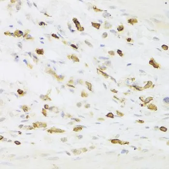
IHC-P analysis of human gastric cancer tissue using GTX33304 LSP1 antibody. Dilution : 1:100


IHC-P analysis of human gastric cancer tissue using GTX33304 LSP1 antibody. Dilution : 1:100
LSP1 antibody
GTX33304
ApplicationsWestern Blot, ImmunoHistoChemistry, ImmunoHistoChemistry Paraffin
Product group Antibodies
ReactivityHuman, Mouse
TargetLSP1
Overview
- SupplierGeneTex
- Product NameLSP1 antibody
- Delivery Days Customer9
- Application Supplier NoteWB: 1:500 - 1:2000. IHC-P: 1:50 - 1:200. *Optimal dilutions/concentrations should be determined by the researcher.Not tested in other applications.
- ApplicationsWestern Blot, ImmunoHistoChemistry, ImmunoHistoChemistry Paraffin
- CertificationResearch Use Only
- ClonalityPolyclonal
- ConjugateUnconjugated
- Gene ID4046
- Target nameLSP1
- Target descriptionlymphocyte specific protein 1
- Target synonymsWP34, pp52, lymphocyte-specific protein 1, 47 kDa actin binding protein, 52 kDa phosphoprotein, F-actin binding and cytoskeleton associated protein, leufactin (leukocyte F-actin binding protein), leukocyte-specific protein 1, lymphocyte-specific antigen WP34
- HostRabbit
- IsotypeIgG
- Protein IDP33241
- Protein NameLymphocyte-specific protein 1
- Scientific DescriptionThis gene encodes an intracellular F-actin binding protein. The protein is expressed in lymphocytes, neutrophils, macrophages, and endothelium and may regulate neutrophil motility, adhesion to fibrinogen matrix proteins, and transendothelial migration. Alternative splicing results in multiple transcript variants encoding different isoforms. [provided by RefSeq, Jul 2008]
- ReactivityHuman, Mouse
- Storage Instruction-20°C or -80°C,2°C to 8°C
- UNSPSC41116161
Datasheet
Related products
Product group Antibodies
Anti-LSP1 AntibodyA101390
ApplicationsWestern Blot, ELISA
ReactivityHuman
- SizePrice

Product group Antibodies
Anti-LSP1 Antibody Picoband(r)A02992-2-CARRIER-FREE
ApplicationsFlow Cytometry, Western Blot, ELISA
ReactivityHuman, Mouse, Rat
TargetLSP1
- SizePrice
Product group Antibodies
Anti-LSP1 Antibody144-05617
ApplicationsWestern Blot, ImmunoHistoChemistry
ReactivityHuman, Mouse
TargetLSP1
- SizePrice
Product group Antibodies

LSP1 AntibodyLS-C748663
ApplicationsWestern Blot
ReactivityHuman, Mouse
TargetLSP1
- SizePrice
Product group Antibodies
LSP1 Recombinant AntibodyBSM-61255R
ApplicationsImmunoFluorescence, Western Blot, ImmunoCytoChemistry, ImmunoHistoChemistry, ImmunoHistoChemistry Frozen, ImmunoHistoChemistry Paraffin
TargetLSP1
- SizePrice
Product group Antibodies
LSP1 AntibodyCSB-PA009880
ApplicationsWestern Blot, ELISA
ReactivityHuman
TargetLSP1
- SizePrice
Product group Antibodies

Goat anti-LSP1 (aa261-275)EB09869
ApplicationsWestern Blot, ELISA
ReactivityBovine, Canine, Human
TargetLSP1
- SizePrice

Product group Antibodies
ApplicationsWestern Blot
ReactivityHuman
TargetLSP1
- SizePrice

Product group Antibodies
LSP1 antibody, C-termGTX88185
ApplicationsWestern Blot, ImmunoHistoChemistry, ImmunoHistoChemistry Paraffin
ReactivityHuman, Mouse, Rat
TargetLSP1
- SizePrice
![Mouse tissue extract (50 μg) was separated by 10% SDS-PAGE, and the membrane was blotted with LSP1 antibody [N2C3] (GTX114533) diluted at 1:1000. The HRP-conjugated anti-rabbit IgG antibody (GTX213110-01) was used to detect the primary antibody.](https://www.genetex.com/upload/website/prouct_img/normal/GTX114533/GTX114533_40499_20180316_WB_M_spleen_w_23060518_230.webp)
Product group Antibodies
LSP1 antibody [N2C3]GTX114533
ApplicationsWestern Blot
ReactivityHuman, Mouse
TargetLSP1
- SizePrice