

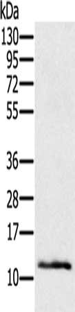
Gel: 12%SDS-PAGE, Lysate: 40 ug, Lane: Mouse skin tissue, Primary antibody: CSB-PA143897(Ly6a Antibody) at dilution 1/200, Secondary antibody: Goat anti rabbit IgG at 1/8000 dilution, Exposure time: 2 minutes
Gel: 12%SDS-PAGE, Lysate: 40 ug, Lane: Mouse skin tissue, Primary antibody: CSB-PA143897(Ly6a Antibody) at dilution 1/200, Secondary antibody: Goat anti rabbit IgG at 1/8000 dilution, Exposure time: 2 minutes
Ly6a Antibody
CSB-PA143897
ApplicationsWestern Blot, ELISA
Product group Antibodies
ReactivityMouse
TargetLy6a
Overview
- SupplierCusabio
- Product NameLy6a Antibody
- Delivery Days Customer20
- ApplicationsWestern Blot, ELISA
- CertificationResearch Use Only
- ClonalityPolyclonal
- ConjugateUnconjugated
- Gene ID110454
- Target nameLy6a
- Target descriptionlymphocyte antigen 6 family member A
- Target synonymsLy-6A.2, Ly-6A/E, Ly-6E.1, Sca-1, Sca1, TAP, lymphocyte antigen 6A-2/6E-1, T-cell-activating protein, lymphocyte antigen 6 complex, locus A, stem cell antigen 1
- HostRabbit
- IsotypeIgG
- Protein IDP05533
- Protein NameLymphocyte antigen 6A-2/6E-1
- Scientific DescriptionSca-1 is an 18 kDa phosphatidylinositol-anchored protein that is a member of the lymphocyte antigen 6 (Ly-6) family. Sca-1 is encoded by the strain-specific Ly-6 E/A allelic gene. Its expression on multipotent hematopoietic stem cells (HSC) has been used as a marker of HSCs in mice of both Ly-6 haplotypes. Sca-1-positive HSCs can be found in the adult bone marrow, fetal liver, and mobilized peripheral blood and spleen in the adult animal.
- ReactivityMouse
- Storage Instruction-20°C or -80°C
- UNSPSC41116161
Related products

Product group Antibodies
Anti-Sca1/Ly6A/E Antibody Picoband(r)A30403-CARRIER-FREE
ApplicationsFlow Cytometry, Western Blot, ImmunoHistoChemistry
ReactivityMouse
TargetLy6a
- SizePrice
Product group Antibodies

Ly6a / Sca-1 AntibodyLS-C662285
ApplicationsWestern Blot
ReactivityMouse
TargetLy6a
- SizePrice
Product group Antibodies
Ly6A Recombinant AntibodyBSM-60617R
ApplicationsImmunoFluorescence, Western Blot, ImmunoHistoChemistry, ImmunoHistoChemistry Frozen, ImmunoHistoChemistry Paraffin
ReactivityMouse
TargetLy6a
- SizePrice
Product group Antibodies
ApplicationsFlow Cytometry
ReactivityMouse
TargetLy6a
- SizePrice