

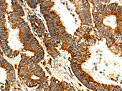
The image on the left is immunohistochemistry of paraffin-embedded Human colon cancer tissue using CSB-PA578056(MAP1LC3A Antibody) at dilution 1/15, on the right is treated with fusion protein. (Original magnification: x200)
 at dilution 1/15, on the right is treated with fusion protein. (Original magnification: x200)")
The image on the left is immunohistochemistry of paraffin-embedded Human colon cancer tissue using CSB-PA578056(MAP1LC3A Antibody) at dilution 1/15, on the right is treated with fusion protein. (Original magnification: x200)
MAP1LC3A Antibody
CSB-PA578056
ApplicationsELISA, ImmunoHistoChemistry
Product group Antibodies
ReactivityHuman, Mouse, Rat
TargetMAP1LC3A
Overview
- SupplierCusabio
- Product NameMAP1LC3A Antibody
- Delivery Days Customer20
- ApplicationsELISA, ImmunoHistoChemistry
- CertificationResearch Use Only
- ClonalityPolyclonal
- ConjugateUnconjugated
- Gene ID84557
- Target nameMAP1LC3A
- Target descriptionmicrotubule associated protein 1 light chain 3 alpha
- Target synonymsATG8E, LC3, LC3A, MAP1ALC3, MAP1BLC3, microtubule-associated protein 1 light chain 3 alpha, MAP1 light chain 3-like protein 1, MAP1A/1B light chain 3 A, MAP1A/MAP1B LC3 A, MAP1A/MAP1B light chain 3 A, autophagy-related ubiquitin-like modifier LC3 A, microtubule-associated proteins 1A/1B light chain 3, microtubule-associated proteins 1A/1B light chain 3A
- HostRabbit
- IsotypeIgG
- Protein IDQ9H492
- Protein NameMicrotubule-associated protein 1 light chain 3 alpha
- Scientific DescriptionMAP1A and MAP1B are microtubule-associated proteins which mediate the physical interactions between microtubules and components of the cytoskeleton. MAP1A and MAP1B each consist of a heavy chain subunit and multiple light chain subunits. The protein encoded by this gene is one of the light chain subunits and can associate with either MAP1A or MAP1B. Two transcript variants encoding different isoforms have been found for this gene. The expression of variant 1 is suppressed in many tumor cell lines, suggesting that may be involved in carcinogenesis.
- ReactivityHuman, Mouse, Rat
- Storage Instruction-20°C or -80°C
- UNSPSC41116161
Related products

Product group Antibodies
Anti-MAP1LC3A Antibody Picoband(r)A01543-1-CARRIER-FREE
ApplicationsWestern Blot, ELISA, ImmunoHistoChemistry
ReactivityHuman, Mouse, Rat
TargetMAP1LC3A
- SizePrice
Product group Antibodies

Cleaved LC3A AntibodyABX029971
ApplicationsImmunoFluorescence, Western Blot, ELISA, ImmunoCytoChemistry, ImmunoHistoChemistry
- SizePrice
Product group Antibodies
Anti-MAP1LC3A AntibodyA30841
ApplicationsImmunoFluorescence, Western Blot, ImmunoHistoChemistry
ReactivityHuman, Mouse, Rat
- SizePrice
Product group Antibodies

MAP1LC3A / LC3A Antibody (clone 7E4)LS-C768002
ApplicationsImmunoFluorescence, Western Blot, ImmunoHistoChemistry, ImmunoHistoChemistry Paraffin
ReactivityHuman, Mouse, Rat
TargetMAP1LC3A
- SizePrice

Product group Antibodies
Anti-MAP1LC3A AntibodyHPA007649
ApplicationsWestern Blot, ImmunoHistoChemistry
ReactivityHuman
TargetMAP1LC3A
- SizePrice
Product group Antibodies
ApplicationsImmunoPrecipitation, Western Blot, ImmunoCytoChemistry, ImmunoHistoChemistry
ReactivityMouse, Porcine, Rat
TargetMAP1LC3A
- SizePrice
Product group Antibodies
LC3A/B Polyclonal AntibodyBS-11731R
ApplicationsImmunoFluorescence, ELISA, ImmunoCytoChemistry, ImmunoHistoChemistry, ImmunoHistoChemistry Frozen, ImmunoHistoChemistry Paraffin
ReactivityBovine, Canine, Chicken, Human, Mouse, Porcine, Rat, Sheep
TargetMAP1LC3A
- SizePrice
Product group Antibodies
Anti-MAP1LC3AY158122
ApplicationsWestern Blot, ELISA, ImmunoHistoChemistry
ReactivityHuman, Mouse, Rat
- SizePrice
Product group Antibodies
LC3A antibodyGTX132889
ApplicationsWestern Blot
ReactivityHuman, Mouse
TargetMAP1LC3A
- SizePrice
Product group Antibodies
Anti-MAP1LC3A Antibody144-05618
ApplicationsImmunoFluorescence, Western Blot, ImmunoHistoChemistry
ReactivityHuman, Mouse, Rat
TargetMAP1LC3A
- SizePrice