

![MART-1 / Melan-A(M2-7C10), CF405S conjugate, 0.1mg/mL [26628-22-8]](https://biotium.com/wp-content/uploads/2016/12/BNUB0009-1-1.jpg "MART-1 / Melan-A(M2-7C10), CF405S conjugate, 0.1mg/mL [26628-22-8]")
MART-1 / Melan-A(M2-7C10), CF405S conjugate, 0.1mg/mL [26628-22-8]
BNC040009
ApplicationsFlow Cytometry, ImmunoFluorescence, Western Blot, ImmunoHistoChemistry, ImmunoHistoChemistry Frozen, ImmunoHistoChemistry Paraffin
Product group Antibodies
ReactivityBovine, Equine, Human, Mouse
TargetMLANA
Overview
- SupplierBiotium
- Product NameMART-1 / Melan-A(M2-7C10), CF405S conjugate, 0.1mg/mL [26628-22-8]
- Delivery Days Customer9
- ApplicationsFlow Cytometry, ImmunoFluorescence, Western Blot, ImmunoHistoChemistry, ImmunoHistoChemistry Frozen, ImmunoHistoChemistry Paraffin
- CAS Number26628-22-8
- CertificationResearch Use Only
- ClonalityMonoclonal
- Clone IDM2-7C10
- Concentration0.1 mg/ml
- ConjugateOther Conjugate
- Gene ID2315
- Target nameMLANA
- Target descriptionmelan-A
- Target synonymsMART-1, MART1, melanoma antigen recognized by T-cells 1, antigen LB39-AA, antigen SK29-AA, epididymis secretory sperm binding protein, protein Melan-A
- HostMouse
- IsotypeIgG2b
- Protein IDQ16655
- Protein NameMelanoma antigen recognized by T-cells 1
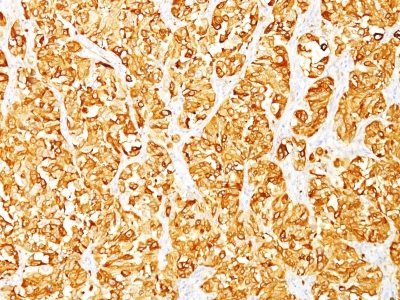
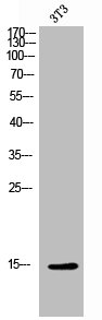
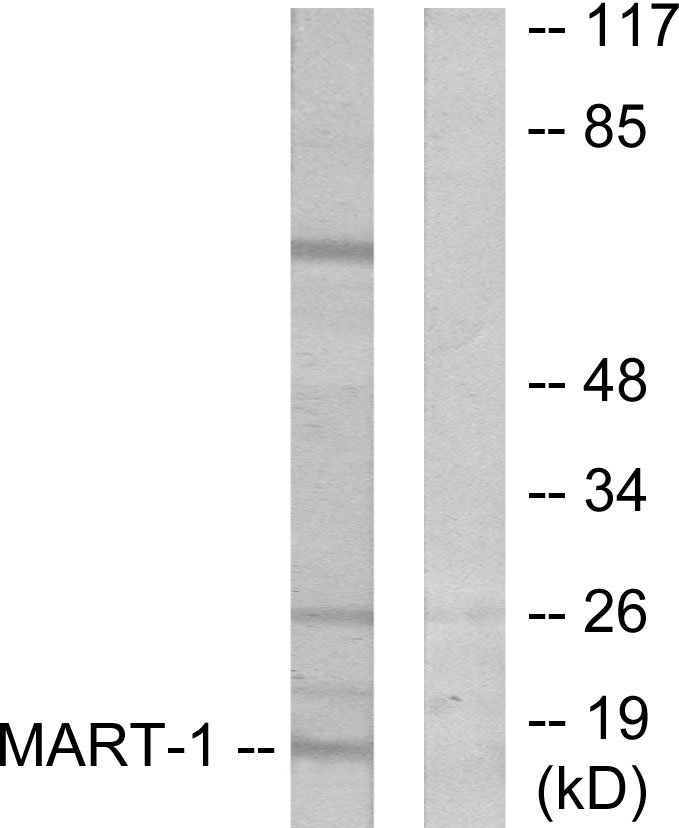
- Scientific DescriptionThis antibody recognizes a protein doublet of 20-22 kDa, identified as MART-1 (Melanoma Antigen Recognized by T cells 1) or Melan-A. MART-1 is a newly identified melanocyte differentiation antigen recognized by autologous cytotoxic T lymphocytes. Seven other melanoma associated antigens recognized by autologous cytotoxic T cells include MAGE-1, MAGE-3, tyrosinase, gp100, gp75, BAGE-1, and GAGE-1. Subcellular fractionation shows that MART-1 is present in melanosomes and endoplasmic reticulum. This MAb labels melanomas and other tumors showing melanocytic differentiation. It is also a useful positive-marker for angiomyolipomas. It does not stain tumor cells of epithelial, lymphoid, glial, or mesenchymal origin.Primary antibodies are available purified, or with a selection of fluorescent CF® Dyes and other labels. CF® Dyes offer exceptional brightness and photostability. Note: Conjugates of blue fluorescent dyes like CF®405S and CF®405M are not recommended for detecting low abundance targets, because blue dyes have lower fluorescence and can give higher non-specific background than other dye colors.
- SourceAnimal
- ReactivityBovine, Equine, Human, Mouse
- Storage Instruction2°C to 8°C,RT
- UNSPSC41116161
MSDS
Related products
Product group Antibodies
MLANA AntibodyCSB-PA008328
ApplicationsWestern Blot, ELISA, ImmunoHistoChemistry
ReactivityHuman
TargetMLANA
- SizePrice
Product group Antibodies
MLANA Polyclonal AntibodyCAC14190
ApplicationsWestern Blot, ELISA, ImmunoHistoChemistry
TargetMLANA
- SizePrice

Product group Antibodies
Anti-Melan-A/MLANA Antibody Picoband(r)A02033-3-CARRIER-FREE
ApplicationsFlow Cytometry, Western Blot, ELISA
ReactivityHuman, Mouse
TargetMLANA
- SizePrice
Product group Antibodies
Anti-MLANA Antibody144-06290
ApplicationsImmunoFluorescence, Western Blot, ImmunoHistoChemistry
ReactivityHuman, Mouse, Rat
TargetMLANA
- SizePrice
Product group Antibodies

MART-1 AntibodyABX013135
ApplicationsWestern Blot, ELISA, ImmunoHistoChemistry
- SizePrice

Product group Antibodies
Anti-MLANA AntibodyAMAB91816
ApplicationsWestern Blot, ImmunoCytoChemistry, ImmunoHistoChemistry
ReactivityHuman
TargetMLANA
- SizePrice
Product group Antibodies

Anti-Melan-A [A103]Ab02840-1.1
ApplicationsImmunoPrecipitation, Western Blot, ELISA, ImmunoHistoChemistry
ReactivityHuman
TargetMLANA
- SizePrice
Product group Antibodies
Anti-MART-1 AntibodyA99292
ApplicationsWestern Blot, ELISA, ImmunoHistoChemistry
ReactivityHuman
- SizePrice
Product group Antibodies

MLANA / Melan-A AntibodyLS-C831121
ApplicationsELISA, ImmunoHistoChemistry
ReactivityHuman
TargetMLANA
- SizePrice