Bio-Connect

Mast Cell Chymase Antibody (YA1629, Rabbit)

HY-P81884
MedChem Express
TargetCMA1
Product group Antibodies
Sign in to order and to see your custom pricing.
Large volume orders?
Order with a bulk request

Overview

  • Supplier
    MedChem Express
  • Product Name
    Mast Cell Chymase Antibody (YA1629, Rabbit)
  • Delivery Days Customer
    5
  • Certification
    Research Use Only
  • Clonality
    Monoclonal
  • Gene ID1215
  • Target name
    CMA1
  • Target description
    chymase 1
  • Target synonyms
    alpha-chymase; chymase; chymase 1 preproprotein transcript E; chymase 1 preproprotein transcript I; chymase 1, mast cell; chymase, heart; chymase, mast cell; CYH; mast cell protease I; MCT1
  • Host
    Rabbit
  • Isotype
    IgG
  • Scientific Description
    Mast Cell Chymase Antibody (YA1629) is a rabbit-derived non-conjugated IgG antibody (Clone NO.: YA1629), targeting Mast Cell Chymase, with a predicted molecular weight of 27 kDa (observed band size: 27 kDa). Mast Cell Chymase Antibody (YA1629) can be used for WB, IHC-P experiment in human background.
  • Storage Instruction
    -20°C
  • UNSPSC
    41116161

Related products

Product group Antibodies
RayBiotech
ApplicationsWestern Blot, ELISA
TargetCMA1
  • SizePrice
Product group Antibodies
Biomatik
ApplicationsWestern Blot, ELISA
TargetCMA1
  • SizePrice
Product group Antibodies
Assay Genie
TargetCMA1
  • SizePrice
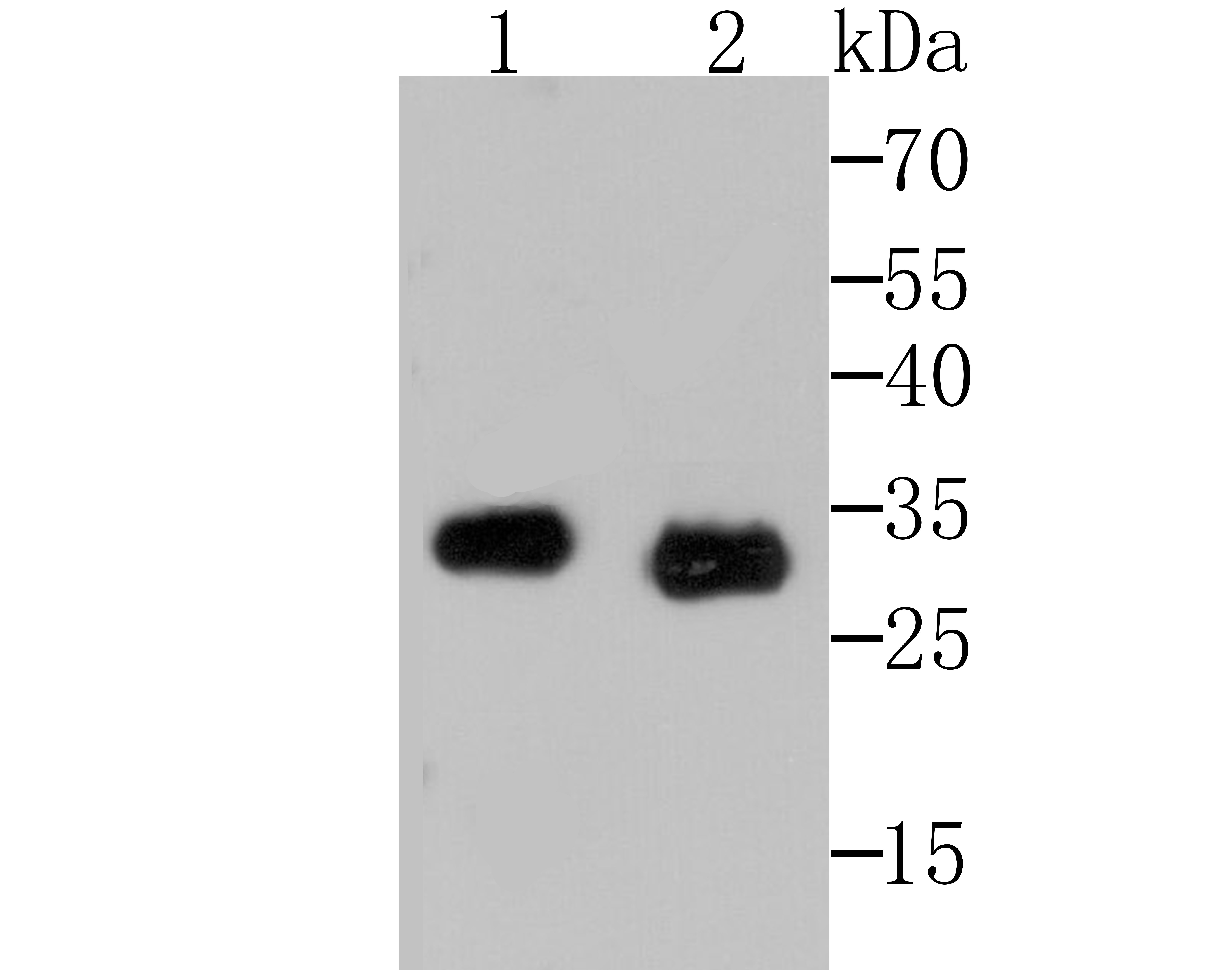
Lane 1: Rat Spleen; Lane 2: Mouse Spleen; Probed with Mast Cell Chymase (1G11) Monoclonal Antibody (bsm-54265R) at 1:500 overnight at 4°C followed by a conjugated secondary antibody for 60 minutes at 37°C.
Product group Antibodies
Bioss Antibodies
ApplicationsImmunoFluorescence, Western Blot, ImmunoHistoChemistry, ImmunoHistoChemistry Frozen, ImmunoHistoChemistry Paraffin
TargetCMA1
  • SizePrice
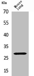
Western Blot analysis of mouse lung cells using Chymase Polyclonal Antibody
Product group Antibodies
Cusabio
CMA1 AntibodyCSB-PA004827
ApplicationsWestern Blot, ELISA
ReactivityHuman
TargetCMA1
  • SizePrice
Immunohistochemical staining of human cerebral cortex shows no positivity in neurons as expected.
Product group Antibodies
Atlas Antibodies
ApplicationsImmunoHistoChemistry
ReactivityHuman
TargetCMA1
  • SizePrice
Product group Antibodies
LifeSpan BioSciences
ApplicationsWestern Blot, ELISA
TargetCMA1
  • SizePrice
Figure 1. IHC analysis of CMA1 using anti-CMA1 antibody (PB9055). CMA1 was detected in a paraffin-embedded section of human placenta tissue. Heat mediated antigen retrieval was performed in EDTA buffer (pH 8.0, epitope retrieval solution). The tissue section was blocked with 10% goat serum. The tissue section was then incubated with 1 microg/ml rabbit anti-CMA1 Antibody (PB9055) overnight at 4°C. Biotinylated goat anti-rabbit IgG was used as secondary antibody and incubated for 30 minutes at 37°C. The tissue section was developed using Strepavidin-Biotin-Complex (SABC) (Catalog # SA1022) with DAB as the chromogen.
Product group Antibodies
Boster Bio
ApplicationsWestern Blot, ImmunoHistoChemistry
TargetCMA1
  • SizePrice
Product group Antibodies
References
GeneTex
ApplicationsWestern Blot, ELISA, ImmunoHistoChemistry, ImmunoHistoChemistry Frozen, ImmunoHistoChemistry Paraffin
TargetCMA1
  • SizePrice
Product group Antibodies
Everest Biotech
ApplicationsWestern Blot, ELISA, ImmunoHistoChemistry
TargetCMA1
  • SizePrice