

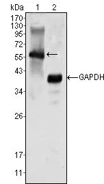
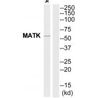
WB analysis of K562 cell lysate (1) using GTX83311 MATK antibody [9D7].
![FACS analysis of K562 cells using GTX83311 MATK antibody [9D7]. Green : MATK Purple : negative control](https://www.genetex.com/upload/website/prouct_img/normal/GTX83311/GTX83311_20170912_FACS_w_23061322_663.webp "FACS analysis of K562 cells using GTX83311 MATK antibody [9D7]. Green : MATK Purple : negative control")
WB analysis of K562 cell lysate (1) using GTX83311 MATK antibody [9D7].
MATK antibody [9D7]
GTX83311
ApplicationsFlow Cytometry, Western Blot, ELISA
Product group Antibodies
ReactivityHuman
TargetMATK
Overview
- SupplierGeneTex
- Product NameMATK antibody [9D7]
- Delivery Days Customer9
- Application Supplier NoteWB: 1/500 - 1/2000. FCM: 1/200 - 1/400. ELISA: 1/10000. *Optimal dilutions/concentrations should be determined by the researcher.Not tested in other applications.
- ApplicationsFlow Cytometry, Western Blot, ELISA
- CertificationResearch Use Only
- ClonalityMonoclonal
- Clone ID9D7
- ConjugateUnconjugated
- Gene ID4145
- Target nameMATK
- Target descriptionmegakaryocyte-associated tyrosine kinase
- Target synonymsCHK, CTK, HHYLTK, HYL, HYLTK, Lsk, megakaryocyte-associated tyrosine-protein kinase, CSK homologous kinase, Csk-homologous kinase, Csk-type protein tyrosine kinase, HYL tyrosine kinase, hematopoietic consensus tyrosine-lacking kinase, hydroxyaryl-protein kinase, leukocyte carboxyl-terminal src kinase related, protein kinase HYL, tyrosine kinase MATK, tyrosine-protein kinase CTK, tyrosylprotein kinase
- HostMouse
- IsotypeIgG1
- Protein IDP42679
- Protein NameMegakaryocyte-associated tyrosine-protein kinase
- Scientific DescriptionThe protein encoded by this gene has amino acid sequence similarity to Csk tyrosine kinase and has the structural features of the CSK subfamily: SRC homology SH2 and SH3 domains, a catalytic domain, a unique N terminus, lack of myristylation signals, lack of a negative regulatory phosphorylation site, and lack of an autophosphorylation site. This protein is thought to play a significant role in the signal transduction of hematopoietic cells. It is able to phosphorylate and inactivate Src family kinases, and may play an inhibitory role in the control of T-cell proliferation. This protein might be involved in signaling in some cases of breast cancer. Three alternatively spliced transcript variants that encode different isoforms have been described for this gene. [provided by RefSeq, Jul 2008]
- ReactivityHuman
- Storage Instruction-20°C or -80°C,2°C to 8°C
- UNSPSC41116161
Datasheet
Related products

Product group Antibodies
Anti-MATK Antibody Picoband(r)A02050-2-CARRIER-FREE
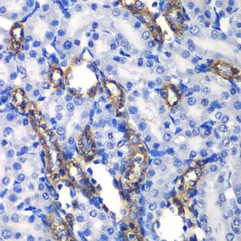
ApplicationsFlow Cytometry, ImmunoFluorescence, Western Blot, ELISA, ImmunoCytoChemistry, ImmunoHistoChemistry
ReactivityHuman, Mouse, Rat
TargetMATK
- SizePrice
Product group Antibodies
Anti-MATK AntibodyA43794
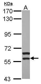
ApplicationsWestern Blot
ReactivityHuman, Mouse, Rat
- SizePrice

Product group Antibodies
Anti-MATK AntibodyHPA004847
ApplicationsWestern Blot, ImmunoHistoChemistry
ReactivityHuman
TargetMATK
- SizePrice
Product group Antibodies
MATK AntibodyCSB-PA010068
ApplicationsWestern Blot, ELISA, ImmunoHistoChemistry
ReactivityHuman, Mouse, Rat
TargetMATK
- SizePrice
Product group Antibodies

MATK AntibodyLS-C334850
ApplicationsWestern Blot, ImmunoHistoChemistry
ReactivityHuman, Mouse, Rat
TargetMATK
- SizePrice
Product group Antibodies
MATK antibodyGTX64561
ApplicationsWestern Blot, ImmunoHistoChemistry, ImmunoHistoChemistry Paraffin
ReactivityHuman, Mouse, Rat
TargetMATK
- SizePrice
Product group Antibodies
MATK antibody [N3C3]GTX111326
ApplicationsWestern Blot
ReactivityHuman
TargetMATK
- SizePrice