

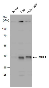
MCL1 antibody detects MCL1 protein by Western blot analysis. Various whole cell extracts (30 μg) were separated by 10% SDS-PAGE, and the membrane was blotted with MCL1 antibody (GTX102026) diluted at 1:1000.
 antibody at 1:250 dilution.
Antigen Retrieval: Trilogy? (EDTA based, pH 8.0) buffer, 15min")
![MCL1 antibody detects MCL1 protein at cytoplasm and nucleus by immunofluorescent analysis. Sample: HeLa cells were fixed in 4% paraformaldehyde at RT for 15 min. Green: MCL1 protein stained by MCL1 antibody (GTX102026) diluted at 1:200. Red: alpha Tubulin, a cytoskeleton marker, stained by alpha Tubulin antibody [B-5-1-2] (GTX11304) diluted at 1:10000. Blue: Hoechst 33342 staining.](https://www.genetex.com/upload/website/prouct_img/normal/GTX102026/GTX102026_39974_20150410_IFA_w_23060100_879.webp "MCL1 antibody detects MCL1 protein at cytoplasm and nucleus by immunofluorescent analysis. Sample: HeLa cells were fixed in 4% paraformaldehyde at RT for 15 min. Green: MCL1 protein stained by MCL1 antibody (GTX102026) diluted at 1:200. Red: alpha Tubulin, a cytoskeleton marker, stained by alpha Tubulin antibody [B-5-1-2] (GTX11304) diluted at 1:10000. Blue: Hoechst 33342 staining.")
 diluted at 1:500.
Antigen Retrieval: Citrate buffer, pH 6.0, 15 min")
 were separated by 10% SDS-PAGE, and the membrane was blotted with MCL1 antibody (GTX102026) diluted at 1:1000. The HRP-conjugated anti-rabbit IgG antibody (GTX213110-01) was used to detect the primary antibody.")

MCL1 antibody detects MCL1 protein by Western blot analysis. Various whole cell extracts (30 μg) were separated by 10% SDS-PAGE, and the membrane was blotted with MCL1 antibody (GTX102026) diluted at 1:1000.
MCL1 antibody
GTX102026
ApplicationsImmunoFluorescence, Western Blot, ImmunoCytoChemistry, ImmunoHistoChemistry, ImmunoHistoChemistry Paraffin
Product group Antibodies
ReactivityHuman, Mouse
TargetMCL1
Overview
- SupplierGeneTex
- Product NameMCL1 antibody
- Delivery Days Customer9
- Application Supplier NoteWB: 1:500-1:3000. ICC/IF: 1:100-1:1000. IHC-P: 1:100-1:1000. *Optimal dilutions/concentrations should be determined by the researcher.Not tested in other applications.
- ApplicationsImmunoFluorescence, Western Blot, ImmunoCytoChemistry, ImmunoHistoChemistry, ImmunoHistoChemistry Paraffin
- CertificationResearch Use Only
- ClonalityPolyclonal
- Concentration0.33 mg/ml
- ConjugateUnconjugated
- Gene ID4170
- Target nameMCL1
- Target descriptionMCL1 apoptosis regulator, BCL2 family member
- Target synonymsBCL2L3, EAT, MCL1-ES, MCL1L, MCL1S, Mcl-1, TM, bcl2-L-3, mcl1/EAT, induced myeloid leukemia cell differentiation protein Mcl-1, BCL2 family apoptosis regulator, MCL1, BCL2 family apoptosis regulator, bcl-2-like protein 3, bcl-2-related protein EAT/mcl1, myeloid cell leukemia 1, myeloid cell leukemia ES, myeloid cell leukemia sequence 1 (BCL2-related)
- HostRabbit
- IsotypeIgG
- Protein IDQ07820
- Protein NameInduced myeloid leukemia cell differentiation protein Mcl-1
- Scientific DescriptionThe protein encoded by this gene belongs to the Bcl-2 family. Alternative splicing occurs at this locus and two transcript variants encoding distinct isoforms have been identified. The longer gene product (isoform 1) enhances cell survival by inhibiting apoptosis while the alternatively spliced shorter gene product (isoform 2) promotes apoptosis and is death-inducing. [provided by RefSeq]
- ReactivityHuman, Mouse
- Storage Instruction-20°C or -80°C,2°C to 8°C
- UNSPSC41116161
Datasheet
Related products
Product group Antibodies
MCL1 AntibodyCSB-PA003207
ApplicationsWestern Blot, ELISA, ImmunoHistoChemistry
ReactivityHuman, Mouse, Rat
TargetMCL1
- SizePrice

Product group Antibodies
Anti-MCL1 Antibody Picoband(r)A00712-2-CARRIER-FREE
ApplicationsFlow Cytometry, ImmunoFluorescence, Western Blot, ELISA, ImmunoCytoChemistry
ReactivityHuman, Mouse, Rat
TargetMCL1
- SizePrice
Product group Antibodies
Anti-MCL1 AntibodyA96164
ApplicationsWestern Blot, ELISA, ImmunoHistoChemistry
ReactivityHuman, Mouse, Rat
- SizePrice

Product group Antibodies
Anti-MCL1 AntibodyAMAB90859
ApplicationsWestern Blot, ImmunoCytoChemistry, ImmunoHistoChemistry
ReactivityHuman
TargetMCL1
- SizePrice
Product group Antibodies

Goat anti-MCL1EB06380
ApplicationsWestern Blot, ELISA
ReactivityBovine, Canine, Human, Mouse, Porcine, Rat
TargetMCL1
- SizePrice
Product group Antibodies

MCL1 / MCL 1 AntibodyLS-C402987
ApplicationsELISA, ImmunoHistoChemistry
ReactivityHuman, Mouse, Rat
TargetMCL1
- SizePrice
Product group Antibodies
MCL1 Polyclonal AntibodyCAC14605
ApplicationsImmunoFluorescence, Western Blot, ELISA, ImmunoHistoChemistry
TargetMCL1
- SizePrice
Product group Antibodies
References
ApplicationsFlow Cytometry, ImmunoFluorescence, Western Blot, ELISA, ImmunoCytoChemistry, ImmunoHistoChemistry, ImmunoHistoChemistry Frozen, ImmunoHistoChemistry Paraffin
ReactivityBovine, Canine, Equine, Human, Mouse, Rat
TargetMCL1
- SizePrice