

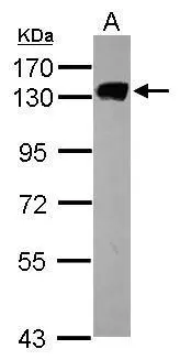
Sample (30 ug of whole cell lysate) A: HCT116 7.5% SDS PAGE GTX103071 diluted at 1:1000
![MCM2 antibody [N1N2], N-term immunoprecipitates MCM2 protein in IP experiments. IP samples: HCT-116 whole cell extract A. 40 μg HCT-116 whole cell extract B. Control with 4 μg of preimmune Rabbit IgG C. Immunoprecipitation of MCM2 protein by 4 μg MCM2 antibody [N1N2], N-term (GTX103071) 7.5 % SDS-PAGE The immunoprecipitated MCM2 protein was detected by MCM2 antibody [N1N2], N-term (GTX103071) diluted at 1:500. [EasyBlot anti-rabbit IgG (GTX221666-01) was used as a secondary reagent]](https://www.genetex.com/upload/website/prouct_img/normal/GTX103071/GTX103071_40450_IP_w_23060119_179.webp "MCM2 antibody [N1N2], N-term immunoprecipitates MCM2 protein in IP experiments. IP samples: HCT-116 whole cell extract A. 40 μg HCT-116 whole cell extract B. Control with 4 μg of preimmune Rabbit IgG C. Immunoprecipitation of MCM2 protein by 4 μg MCM2 antibody [N1N2], N-term (GTX103071) 7.5 % SDS-PAGE The immunoprecipitated MCM2 protein was detected by MCM2 antibody [N1N2], N-term (GTX103071) diluted at 1:500. [EasyBlot anti-rabbit IgG (GTX221666-01) was used as a secondary reagent]")
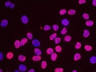
![MCM2 antibody [N1N2], N-term detects MCM2 protein at nucleus by immunofluorescent analysis. Sample: HCT116 cells were fixed in 4% paraformaldehyde at RT for 15 min. Green: MCM2 protein stained by MCM2 antibody [N1N2], N-term (GTX103071) diluted at 1:500. Red: phalloidin, a cytoskeleton marker, diluted at 1:50.](https://www.genetex.com/upload/website/prouct_img/normal/GTX103071/GTX103071_40450_20160629_IFA_w_23060119_855.webp "MCM2 antibody [N1N2], N-term detects MCM2 protein at nucleus by immunofluorescent analysis. Sample: HCT116 cells were fixed in 4% paraformaldehyde at RT for 15 min. Green: MCM2 protein stained by MCM2 antibody [N1N2], N-term (GTX103071) diluted at 1:500. Red: phalloidin, a cytoskeleton marker, diluted at 1:50.")
Sample (30 ug of whole cell lysate) A: HCT116 7.5% SDS PAGE GTX103071 diluted at 1:1000
MCM2 antibody [N1N2], N-term
GTX103071
ApplicationsImmunoFluorescence, ImmunoPrecipitation, Western Blot, ImmunoCytoChemistry
Product group Antibodies
ReactivityHuman
TargetMCM2
Overview
- SupplierGeneTex
- Product NameMCM2 antibody [N1N2], N-term
- Delivery Days Customer9
- Application Supplier NoteWB: 1:500-1:3000. ICC/IF: 1:100-1:1000. IP: 1:100-1:500. *Optimal dilutions/concentrations should be determined by the researcher.Not tested in other applications.
- ApplicationsImmunoFluorescence, ImmunoPrecipitation, Western Blot, ImmunoCytoChemistry
- CertificationResearch Use Only
- ClonalityPolyclonal
- Concentration1 mg/ml
- ConjugateUnconjugated
- Gene ID4171
- Target nameMCM2
- Target descriptionminichromosome maintenance complex component 2
- Target synonymsBM28, CCNL1, CDCL1, D3S3194, DFNA70, MITOTIN, cdc19, DNA replication licensing factor MCM2, cell devision cycle-like 1, cyclin-like 1, minichromosome maintenance deficient 2 (mitotin), minichromosome maintenance protein 2 homolog, nuclear protein BM28
- HostRabbit
- IsotypeIgG
- Protein IDP49736
- Protein NameDNA replication licensing factor MCM2
- Scientific DescriptionThe protein encoded by this gene is one of the highly conserved mini-chromosome maintenance proteins (MCM) that are involved in the initiation of eukaryotic genome replication. The hexameric protein complex formed by MCM proteins is a key component of the pre-replication complex (pre_RC) and may be involved in the formation of replication forks and in the recruitment of other DNA replication related proteins. This protein forms a complex with MCM4, 6, and 7, and has been shown to regulate the helicase activity of the complex. This protein is phosphorylated, and thus regulated by, protein kinases CDC2 and CDC7. [provided by RefSeq]
- ReactivityHuman
- Storage Instruction-20°C or -80°C,2°C to 8°C
- UNSPSC41116161
Datasheet
Related products
Product group Antibodies
Anti-MCM2 AntibodyA96383
ApplicationsImmunoFluorescence, Western Blot, ELISA, ImmunoHistoChemistry
ReactivityHuman, Mouse
- SizePrice
Product group Antibodies
Anti-MCM2 Antibody144-01056
ApplicationsWestern Blot, ImmunoHistoChemistry
ReactivityHuman, Mouse, Rat
TargetMCM2
- SizePrice

Product group Antibodies
Anti-MCM2 Antibody Picoband(r)A00374-1-CARRIER-FREE
ApplicationsFlow Cytometry, ImmunoFluorescence, Western Blot, ELISA, ImmunoCytoChemistry, ImmunoHistoChemistry
ReactivityHuman, Mouse, Rat
TargetMCM2
- SizePrice
Product group Antibodies
ApplicationsImmunoFluorescence, ELISA, ImmunoCytoChemistry, ImmunoHistoChemistry, ImmunoHistoChemistry Frozen, ImmunoHistoChemistry Paraffin
ReactivityCanine, Chicken, Equine, Human, Mouse, Porcine, Rabbit, Rat, Sheep, Zebra Fish
TargetMCM2
- SizePrice
Product group Antibodies
MCM2 AntibodyCSB-PA000999
ApplicationsImmunoFluorescence, Western Blot, ELISA, ImmunoHistoChemistry
ReactivityHuman, Mouse
TargetMCM2
- SizePrice
Product group Antibodies
MCM2 Polyclonal AntibodyCAC13821
ApplicationsImmunoFluorescence, Western Blot, ELISA, ImmunoHistoChemistry
TargetMCM2
- SizePrice
Product group Antibodies
MCM2 antibodyGTX24461
ApplicationsImmunoPrecipitation, Western Blot, ImmunoHistoChemistry, ImmunoHistoChemistry Paraffin, Other Application
ReactivityHuman, Xenopus
TargetMCM2
- SizePrice
![IHC-P analysis of human colonic crypts tissue using GTX04968 MCM2 antibody [MSVA-502R] HistoMAX?. MCM2 staining is strongest in epithelial cells of colonic crypts. Some lymphocytes are also positive scaled.](https://www.genetex.com/upload/website/prouct_img/normal/GTX04968/GTX04968_20241028_IHC-P_24102820_901.webp)
Product group Antibodies
ApplicationsImmunoHistoChemistry, ImmunoHistoChemistry Paraffin
ReactivityHuman
TargetMCM2
- SizePrice
![IHC-P analysis of human tonsil tissue using GTX75459 MCM2 antibody [CRCT2.1 (D1.9H5)].](https://www.genetex.com/upload/website/prouct_img/normal/GTX75459/GTX75459_4902_IHC-P_w_23051501_670.webp)
Product group Antibodies
MCM2 antibody [CRCT2.1 (D1.9H5)]GTX75459
ApplicationsWestern Blot, ImmunoHistoChemistry, ImmunoHistoChemistry Paraffin
ReactivityHuman
TargetMCM2
- SizePrice
![IHC-P analysis of colon cancer tissue using GTX60491 MCM2 antibody [2B3].](https://www.genetex.com/upload/website/prouct_img/normal/GTX60491/GTX60491_20170912_IHC-P_w_23061123_117.webp)
Product group Antibodies
MCM2 antibody [2B3]GTX60491
ApplicationsFlow Cytometry, Western Blot, ELISA, ImmunoHistoChemistry, ImmunoHistoChemistry Paraffin
ReactivityHuman, Monkey, Mouse, Rat
TargetMCM2
- SizePrice