

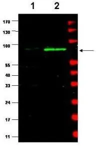
Western blot using GeneTex's affinity purified Anti-MDM2 (Rabbit) is shown to detect a band (arrow) corresponding to mouse MDM2 protein present in mouse MEF cells (lane 2) but not human kidney HEK293 cells (lane 1). Approximately 35 ug of lysate was separated by 4-20% Tris Glycine SDS-PAGE. After blocking the membrane with 5% normal goat serum, 0.5% BLOTTO in PBS, the membrane was probed for overnight at 4o with the primary antibody diluted to 1:500 in 1% normal goat serum, 0.1% BLOTTO in PBS. The membrane was washed and reacted with a 1:10,000 dilution of IRDye800 conjugated Gt-a-Rabbit IgG [H&L] for 45 min at room temperature (800 nm channel, green). Molecular weight estimation was made by comparison to prestained MW markers indicated at the right (700 nm channel, red). IRDye800 fluorescence image was captured using the OdysseyR Infrared Imaging System developed by LI-COR. IRDye is a trademark of LI-COR, Inc. Other detection systems will yield similar results.

Western blot using GeneTex's affinity purified Anti-MDM2 (Rabbit) is shown to detect a band (arrow) corresponding to mouse MDM2 protein present in mouse MEF cells (lane 2) but not human kidney HEK293 cells (lane 1). Approximately 35 ug of lysate was separated by 4-20% Tris Glycine SDS-PAGE. After blocking the membrane with 5% normal goat serum, 0.5% BLOTTO in PBS, the membrane was probed for overnight at 4o with the primary antibody diluted to 1:500 in 1% normal goat serum, 0.1% BLOTTO in PBS. The membrane was washed and reacted with a 1:10,000 dilution of IRDye800 conjugated Gt-a-Rabbit IgG [H&L] for 45 min at room temperature (800 nm channel, green). Molecular weight estimation was made by comparison to prestained MW markers indicated at the right (700 nm channel, red). IRDye800 fluorescence image was captured using the OdysseyR Infrared Imaging System developed by LI-COR. IRDye is a trademark of LI-COR, Inc. Other detection systems will yield similar results.
MDM2 antibody
GTX48728
ApplicationsImmunoPrecipitation, Western Blot, ELISA
Product group Antibodies
ReactivityMouse
TargetMdm2
Overview
- SupplierGeneTex
- Product NameMDM2 antibody
- Delivery Days Customer9
- Application Supplier NoteWB: 1:500-1:2000. IP: 1:100. ELISA: 1:3000-1:12000. *Optimal dilutions/concentrations should be determined by the researcher.Not tested in other applications.
- ApplicationsImmunoPrecipitation, Western Blot, ELISA
- CertificationResearch Use Only
- ClonalityPolyclonal
- Concentration1 mg/ml
- ConjugateUnconjugated
- Gene ID17246
- Target nameMdm2
- Target descriptiontransformed mouse 3T3 cell double minute 2
- Target synonyms1700007J15Rik, Mdm-2, E3 ubiquitin-protein ligase Mdm2, RING-type E3 ubiquitin transferase Mdm2, double minute 2 protein, oncoprotein Mdm2, p53-binding protein Mdm2
- HostRabbit
- IsotypeIgG
- Protein IDP23804
- Protein NameE3 ubiquitin-protein ligase Mdm2
- Scientific DescriptionMDM2 is a nuclear phosphoprotein with an apparent molecular mass of 90 kD that forms a complex with the p53 tumor suppressor protein. Human MDM2 was identified as a homologous product of the murine double minute 2 gene (mdm2). The MDM2 gene enhances the tumorigenic potential of cells when it is overexpressed and encodes a putative transcription factor. Forming a tight complex with the p53 gene, the MDM2 oncogene can inhibit p53-mediated transactivation. MDM2 binds to p53 and amplification of MDM2 in sarcomas leads to escape from p53-regulated growth control. This mechanism of tumorigenesis parallels that for virus-induced tumors in which viral oncogene products bind to and functionally inactivate p53.Over-expression of the
- ReactivityMouse
- Storage Instruction-20°C or -80°C,2°C to 8°C
- UNSPSC41116161
Datasheet
Related products
Product group Antibodies
Mdm2 AntibodyCSB-PA013626ZA01MO
ApplicationsWestern Blot, ELISA
ReactivityMouse
TargetMdm2
- SizePrice
Product group Antibodies
MDM2 antibody [MD-219]GTX10344
ApplicationsFlow Cytometry, ImmunoFluorescence, Western Blot, ELISA, ImmunoCytoChemistry
ReactivityMouse
TargetMdm2
- SizePrice
Product group Antibodies
MDM2 (phospho Ser185) antibodyGTX21094
ApplicationsImmunoPrecipitation, Western Blot, ELISA
ReactivityHuman, Mouse
TargetMdm2
- SizePrice