

Affinity Purified Anti-MDM2 pS185 (Rabbit) is shown to detect a 102 kDa band (arrow) corresponding to phosphorylated mouse MDM2 present in a 293T whole cell lysate. Cells were serum-starved for 24 hours prior to harvest. Approxi-mately 20 ug of lysate was loaded per lane for SDS-PAGE. Untreated cells are shown in lane 1, whereas cells in lane 2 were treated with IGF-1 (100 ng/ml) for 20 min prior to harvest. Follow reaction of antibody with a 1:2000 dilution of HRP Goat-a-Rabbit IgG for visualization.
 antibody. Lane 1 : Untreated whole cell lysate Lane 2 : 100 ng/mL IGF-1 treated whole cell lysate Dilution : 20 μg")
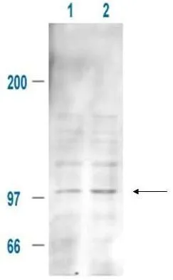
Affinity Purified Anti-MDM2 pS185 (Rabbit) is shown to detect a 102 kDa band (arrow) corresponding to phosphorylated mouse MDM2 present in a 293T whole cell lysate. Cells were serum-starved for 24 hours prior to harvest. Approxi-mately 20 ug of lysate was loaded per lane for SDS-PAGE. Untreated cells are shown in lane 1, whereas cells in lane 2 were treated with IGF-1 (100 ng/ml) for 20 min prior to harvest. Follow reaction of antibody with a 1:2000 dilution of HRP Goat-a-Rabbit IgG for visualization.
MDM2 (phospho Ser185) antibody
GTX21094
ApplicationsImmunoPrecipitation, Western Blot, ELISA
Product group Antibodies
ReactivityHuman, Mouse
TargetMdm2
Overview
- SupplierGeneTex
- Product NameMDM2 (phospho Ser185) antibody
- Delivery Days Customer9
- Application Supplier NoteWB: 1:500-1:2000. IP: 1:100. ELISA: 1:3000-1:12000. *Optimal dilutions/concentrations should be determined by the researcher.Not tested in other applications.
- ApplicationsImmunoPrecipitation, Western Blot, ELISA
- CertificationResearch Use Only
- ClonalityPolyclonal
- Concentration1 mg/ml
- ConjugateUnconjugated
- Gene ID17246
- Target nameMdm2
- Target descriptiontransformed mouse 3T3 cell double minute 2
- Target synonyms1700007J15Rik, Mdm-2, E3 ubiquitin-protein ligase Mdm2, RING-type E3 ubiquitin transferase Mdm2, double minute 2 protein, oncoprotein Mdm2, p53-binding protein Mdm2
- HostRabbit
- IsotypeIgG
- Protein IDP23804
- Protein NameE3 ubiquitin-protein ligase Mdm2
- Scientific DescriptionMDM2 is a nuclear phosphoprotein with an apparent molecular mass of 90 Kd that forms a complex with the p53 tumor suppressor protein. Human MDM2 was identified as a homologous product of the murine double minute 2 gene (mdm2). The MDM2 gene enhances the tumorigenic potential of cells when it is overexpressed and encodes a putative transcription factor. Forming a tight complex with the p53 gene, the MDM2 oncogene can inhibit p53-mediated transactivation. MDM2 binds to p53 and amplification of MDM2 in sarcomas leads to escape from p53-regulated growth control. This mechanism of tumorigenesis parallels that for virus-induced tumors in which viral oncogene products bind to and functionally inactivate p53. Overexpression of the MDM2 oncogene was found in leukemias. Inactivation of tumor suppressor genes leads to deregulated cell proliferation and is a key factor in human tumorigenesis. MDM2 interacts physically and functionally with the retinoblastoma (RB) protein and can inhibit its growth regulatory capacity. Both RB and p53 can be subjected to negative regulation by the product of a single cellular protooncogene. The interference of binding to p53 prevents the interaction of MDM2 and its regulation of the transcriptional activity of p53 in vivo. Direct association of p53 with the cellular protein MDM2 results in ubiquitination and subsequent degradation of p53. MDM2-p53 complexes were preferentially found in S/G2M phases of the cell cycle. MDM2 maps to 12q14.3-q15, distal to CDK4 and flanked by Genethon microsatellites D12S80 and D12S83. On both the physical and the genetic maps of chromosome 12, the IFG gene maps close to the locus of the MDM2 oncogene on 12q15. The MDM2 gene is alternatively spliced, producing 5 additional splice variant transcripts from the full length MDM2 gene. Four out of five of these alternatively spliced forms (MDM2a-MDMd) are missing substantial portions of the p53-binding domain and retain the acidic domain and the zinc-finger domains. The fifth and smallest transcript (MDM2e) retains the largest spliced region encoding the p53-binding domain; however, it lacks the nuclear localization signal, the acidic domain and zinc-finger domains. The alternatively spliced transcripts tend to be expressed in tumorigenic tissue, whereas the full length MDM2 transcript is expressed in normal tissue.
- ReactivityHuman, Mouse
- Storage Instruction-20°C or -80°C,2°C to 8°C
- UNSPSC41116161
Datasheet
Related products
Product group Antibodies
Mdm2 AntibodyCSB-PA013626ZA01MO
ApplicationsWestern Blot, ELISA
ReactivityMouse
TargetMdm2
- SizePrice
Product group Antibodies
MDM2 antibody [MD-219]GTX10344
ApplicationsFlow Cytometry, ImmunoFluorescence, Western Blot, ELISA, ImmunoCytoChemistry
ReactivityMouse
TargetMdm2
- SizePrice
![Western blot using GeneTex's affinity purified Anti-MDM2 (Rabbit) is shown to detect a band (arrow) corresponding to mouse MDM2 protein present in mouse MEF cells (lane 2) but not human kidney HEK293 cells (lane 1). Approximately 35 ug of lysate was separated by 4-20% Tris Glycine SDS-PAGE. After blocking the membrane with 5% normal goat serum, 0.5% BLOTTO in PBS, the membrane was probed for overnight at 4o with the primary antibody diluted to 1:500 in 1% normal goat serum, 0.1% BLOTTO in PBS. The membrane was washed and reacted with a 1:10,000 dilution of IRDye800 conjugated Gt-a-Rabbit IgG [H&L] for 45 min at room temperature (800 nm channel, green). Molecular weight estimation was made by comparison to prestained MW markers indicated at the right (700 nm channel, red). IRDye800 fluorescence image was captured using the OdysseyR Infrared Imaging System developed by LI-COR. IRDye is a trademark of LI-COR, Inc. Other detection systems will yield similar results.](https://www.genetex.com/upload/website/prouct_img/normal/GTX48728/GTX48728_20160330_WB_w_23060823_413.webp)
Product group Antibodies
MDM2 antibodyGTX48728
ApplicationsImmunoPrecipitation, Western Blot, ELISA
ReactivityMouse
TargetMdm2
- SizePrice