

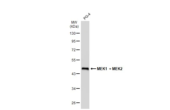
Whole cell extract (30 μg) was separated by 10% SDS-PAGE, and the membrane was blotted with MEK1 + MEK2 antibody [GT1721] (GTX630542) diluted at 1:5000. The HRP-conjugated anti-mouse IgG antibody (GTX213111-01) was used to detect the primary antibody.
![Whole cell extract (30 μg) was separated by 10% SDS-PAGE, and the membrane was blotted with MEK1 + MEK2 antibody [GT1721] (GTX630542) diluted at 1:5000. The HRP-conjugated anti-mouse IgG antibody (GTX213111-01) was used to detect the primary antibody.](https://www.genetex.com/upload/website/prouct_img/normal/GTX630542/GTX630542_41568_20190222_WB_D_22111423_501.webp "Whole cell extract (30 μg) was separated by 10% SDS-PAGE, and the membrane was blotted with MEK1 + MEK2 antibody [GT1721] (GTX630542) diluted at 1:5000. The HRP-conjugated anti-mouse IgG antibody (GTX213111-01) was used to detect the primary antibody.")
![MEK1 + MEK2 antibody [GT1721] detects MEK1 + MEK2 protein at cytoplasm by immunofluorescent analysis. Sample: MDCK cells were fixed in 4% paraformaldehyde at RT for 15 min. Green: MEK2 stained by MEK1 + MEK2 antibody [GT1721] (GTX630542) diluted at 1:500. Blue: Hoechst 33342 staining.](https://www.genetex.com/upload/website/prouct_img/normal/GTX630542/GTX630542_41568_20190612_ICC_IF_D_22111423_387.webp "MEK1 + MEK2 antibody [GT1721] detects MEK1 + MEK2 protein at cytoplasm by immunofluorescent analysis. Sample: MDCK cells were fixed in 4% paraformaldehyde at RT for 15 min. Green: MEK2 stained by MEK1 + MEK2 antibody [GT1721] (GTX630542) diluted at 1:500. Blue: Hoechst 33342 staining.")
![Whole cell extract (30 μg) was separated by 10% SDS-PAGE, and the membrane was blotted with MEK1 + MEK2 antibody [GT1721] (GTX630542) diluted at 1:5000. The HRP-conjugated anti-mouse IgG antibody (GTX213111-01) was used to detect the primary antibody, and the signal was developed with Trident ECL plus-Enhanced.](https://www.genetex.com/upload/website/prouct_img/normal/GTX630542/GTX630542_41568_20230210_WB_Drosophila_23021401_335.webp "Whole cell extract (30 μg) was separated by 10% SDS-PAGE, and the membrane was blotted with MEK1 + MEK2 antibody [GT1721] (GTX630542) diluted at 1:5000. The HRP-conjugated anti-mouse IgG antibody (GTX213111-01) was used to detect the primary antibody, and the signal was developed with Trident ECL plus-Enhanced.")
![MEK2 antibody [GT1721] detects MEK2 protein by western blot analysis. A. 30 μg 293T whole cell lysate/extract B. 30 μg A431 whole cell lysate/extract C. 30 μg HeLa whole cell lysate/extract | D. 30 μg HepG2 whole cell lysate/extract 10 % SDS-PAGE MEK2 antibody [GT1721] (GTX630542) dilution: 1:5000](https://www.genetex.com/upload/website/prouct_img/normal/GTX630542/GTX630542_41568_WB_w_23061202_534.webp "MEK2 antibody [GT1721] detects MEK2 protein by western blot analysis. A. 30 μg 293T whole cell lysate/extract B. 30 μg A431 whole cell lysate/extract C. 30 μg HeLa whole cell lysate/extract | D. 30 μg HepG2 whole cell lysate/extract 10 % SDS-PAGE MEK2 antibody [GT1721] (GTX630542) dilution: 1:5000")
![MEK1 + MEK2 antibody [GT1721] detects MEK1 + MEK2 protein at cytoplasm by immunofluorescent analysis. Sample: HeLa cells were fixed in 4% paraformaldehyde at RT for 15 min. Green: MEK1 + MEK2 stained by MEK1 + MEK2 antibody [GT1721] (GTX630542) diluted at 1:500. Blue: Hoechst 33342 staining.](https://www.genetex.com/upload/website/prouct_img/normal/GTX630542/GTX630542_41568_20190703_ICC_IF_w_23061202_540.webp "MEK1 + MEK2 antibody [GT1721] detects MEK1 + MEK2 protein at cytoplasm by immunofluorescent analysis. Sample: HeLa cells were fixed in 4% paraformaldehyde at RT for 15 min. Green: MEK1 + MEK2 stained by MEK1 + MEK2 antibody [GT1721] (GTX630542) diluted at 1:500. Blue: Hoechst 33342 staining.")
Whole cell extract (30 μg) was separated by 10% SDS-PAGE, and the membrane was blotted with MEK1 + MEK2 antibody [GT1721] (GTX630542) diluted at 1:5000. The HRP-conjugated anti-mouse IgG antibody (GTX213111-01) was used to detect the primary antibody.
MEK1 + MEK2 antibody [GT1721]
GTX630542
ApplicationsImmunoFluorescence, Western Blot, ImmunoCytoChemistry
Product group Antibodies
ReactivityCanine, Drosophila, Feline, Human
Overview
- SupplierGeneTex
- Product NameMEK1 + MEK2 antibody [GT1721]
- Delivery Days Customer9
- Application Supplier NoteWB: 1:1000-1:10000. *Optimal dilutions/concentrations should be determined by the researcher.Not tested in other applications.
- ApplicationsImmunoFluorescence, Western Blot, ImmunoCytoChemistry
- CertificationResearch Use Only
- ClonalityMonoclonal
- Clone IDGT1721
- Concentration1 mg/ml
- ConjugateUnconjugated
- HostMouse
- IsotypeIgG1
- ReactivityCanine, Drosophila, Feline, Human
- Storage Instruction-20°C or -80°C,2°C to 8°C
- UNSPSC41116161