

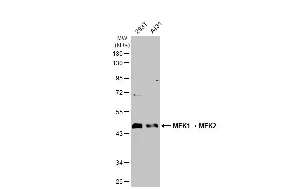
Various whole cell extracts (30 μg) were separated by 10% SDS-PAGE, and the membrane was blotted with MEK1 + MEK2 antibody (GTX121942) diluted at 1:1000. The HRP-conjugated anti-rabbit IgG antibody (GTX213110-01) was used to detect the primary antibody.
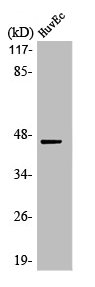
 was separated by 10% SDS-PAGE, and the membrane was blotted with MEK1 + MEK2 antibody (GTX121942) diluted at 1:3000. The HRP-conjugated anti-rabbit IgG antibody (GTX213110-01) was used to detect the primary antibody, and the signal was developed with Trident ECL plus-Enhanced.")
 diluted at 1:500. Blue: Hoechst 33342 staining.")
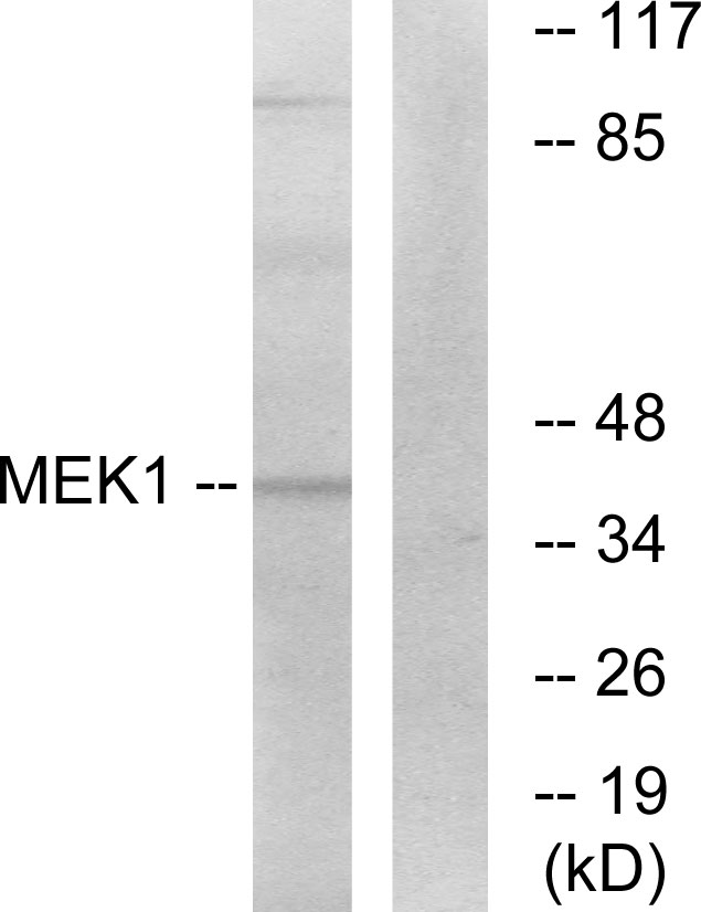
 A: NIH-3T3 B: JC 10% SDS PAGE GTX121942 diluted at 1:2000")
 diluted at 1:500. Blue: Hoechst 33342 staining.")
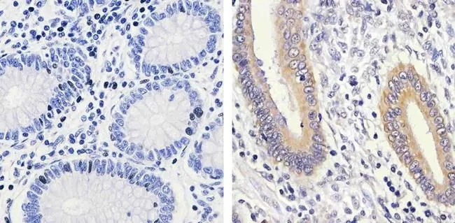
 diluted at 1:500.
Antigen Retrieval: Citrate buffer, pH 6.0, 15 min")
Various whole cell extracts (30 μg) were separated by 10% SDS-PAGE, and the membrane was blotted with MEK1 + MEK2 antibody (GTX121942) diluted at 1:1000. The HRP-conjugated anti-rabbit IgG antibody (GTX213110-01) was used to detect the primary antibody.
MEK1 + MEK2 antibody
GTX121942
ApplicationsImmunoFluorescence, Western Blot, ImmunoCytoChemistry, ImmunoHistoChemistry, ImmunoHistoChemistry Paraffin
Product group Antibodies
ReactivityCanine, Human, Mouse, Rat
TargetMAP2K1
Overview
- SupplierGeneTex
- Product NameMEK1 + MEK2 antibody
- Delivery Days Customer9
- Application Supplier NoteWB: 1:500-1:3000. ICC/IF: 1:100-1:1000. IHC-P: 1:100-1:1000. *Optimal dilutions/concentrations should be determined by the researcher.Not tested in other applications.
- ApplicationsImmunoFluorescence, Western Blot, ImmunoCytoChemistry, ImmunoHistoChemistry, ImmunoHistoChemistry Paraffin
- CertificationResearch Use Only
- ClonalityPolyclonal
- Concentration1 mg/ml
- ConjugateUnconjugated
- Gene ID5604
- Target nameMAP2K1
- Target descriptionmitogen-activated protein kinase kinase 1
- Target synonymsCFC3, MAPKK1, MEK1, MEL, MKK1, PRKMK1, dual specificity mitogen-activated protein kinase kinase 1, ERK activator kinase 1, MAPK/ERK kinase 1, MAPKK 1, MEK 1, protein kinase, mitogen-activated, kinase 1 (MAP kinase kinase 1)
- HostRabbit
- IsotypeIgG
- ReactivityCanine, Human, Mouse, Rat
- Storage Instruction-20°C or -80°C,2°C to 8°C
- UNSPSC41116161
Datasheet
Related products
Product group Antibodies
MAP2K1 AntibodyCSB-PA003217
ApplicationsWestern Blot, ELISA, ImmunoHistoChemistry
ReactivityHuman, Mouse, Rat
TargetMAP2K1
- SizePrice

Product group Antibodies
Anti-MEK1/MAP2K1 Antibody Picoband(r)A00292-CARRIER-FREE
ApplicationsFlow Cytometry, ImmunoFluorescence, Western Blot, ELISA, ImmunoCytoChemistry
ReactivityHuman, Mouse, Rat
TargetMAP2K1
- SizePrice
Product group Antibodies
Anti-MEK1 AntibodyA96159
ApplicationsWestern Blot, ELISA, ImmunoHistoChemistry
ReactivityHuman, Mouse, Rat
- SizePrice

Product group Antibodies
Anti-MAP2K1 AntibodyHPA026430
ApplicationsWestern Blot, ImmunoCytoChemistry, ImmunoHistoChemistry
ReactivityHuman
TargetMAP2K1
- SizePrice
Product group Antibodies

MAP2K1 / MKK1 / MEK1 AntibodyLS-C405977
ApplicationsWestern Blot, ELISA, ImmunoHistoChemistry
ReactivityHuman, Mouse, Rat
TargetMAP2K1
- SizePrice
Product group Antibodies
MEK1 (phospho Thr292) antibodyGTX25612
ApplicationsFlow Cytometry, ImmunoFluorescence, Western Blot, ImmunoCytoChemistry, ImmunoHistoChemistry, ImmunoHistoChemistry Paraffin
ReactivityHuman, Mouse, Rat
TargetMAP2K1
- SizePrice
Product group Antibodies
References
MEK1 (phospho Ser298) antibodyGTX25613
ApplicationsFlow Cytometry, ImmunoFluorescence, Western Blot, ImmunoCytoChemistry, ImmunoHistoChemistry, ImmunoHistoChemistry Paraffin
ReactivityHuman, Mouse, Rat
TargetMAP2K1
- SizePrice
Product group Antibodies
MAP2K1 Polyclonal AntibodyCAC14738
ApplicationsWestern Blot, ELISA, ImmunoHistoChemistry
ReactivityMouse
TargetMAP2K1
- SizePrice