

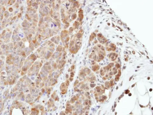
Immunohistochemical analysis of paraffin-embedded SW480 xenograft, using MEK3(GTX107529) antibody at 1:500 dilution.
Antigen Retrieval: Citrate buffer, pH 6.0, 15 min
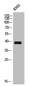
 A: mouse liver 10% SDS PAGE GTX107529 diluted at 1:500")
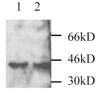
![Various whole cell extracts (30 μg) were separated by 10% SDS-PAGE, and the membrane was blotted with MEK3 antibody [N1C3] (GTX107529) diluted at 1:1000. The HRP-conjugated anti-rabbit IgG antibody (GTX213110-01) was used to detect the primary antibody.](https://www.genetex.com/upload/website/prouct_img/normal/GTX107529/GTX107529_40037_20220311_WB_w_23060120_852.webp "Various whole cell extracts (30 μg) were separated by 10% SDS-PAGE, and the membrane was blotted with MEK3 antibody [N1C3] (GTX107529) diluted at 1:1000. The HRP-conjugated anti-rabbit IgG antibody (GTX213110-01) was used to detect the primary antibody.")
 antibody at 1:200 dilution.")
![MEK3 antibody [N1C3] detects MEK3 protein at cytoplasm in mouse colon by immunohistochemical analysis. Sample: Paraffin-embedded mouse colon. MEK3 antibody [N1C3] (GTX107529) diluted at 1:500.
Antigen Retrieval: Citrate buffer, pH 6.0, 15 min](https://www.genetex.com/upload/website/prouct_img/normal/GTX107529/GTX107529_40037_20150420_IHC_M_2_w_23060120_953.webp "MEK3 antibody [N1C3] detects MEK3 protein at cytoplasm in mouse colon by immunohistochemical analysis. Sample: Paraffin-embedded mouse colon. MEK3 antibody [N1C3] (GTX107529) diluted at 1:500.
Antigen Retrieval: Citrate buffer, pH 6.0, 15 min")
![Various whole cell extracts (30 μg) were separated by 10% SDS-PAGE, and the membrane was blotted with MEK3 antibody [N1C3] (GTX107529) diluted at 1:1000. The HRP-conjugated anti-rabbit IgG antibody (GTX213110-01) was used to detect the primary antibody.](https://www.genetex.com/upload/website/prouct_img/normal/GTX107529/GTX107529_40037_20220311_WB_2_w_23060120_102.webp "Various whole cell extracts (30 μg) were separated by 10% SDS-PAGE, and the membrane was blotted with MEK3 antibody [N1C3] (GTX107529) diluted at 1:1000. The HRP-conjugated anti-rabbit IgG antibody (GTX213110-01) was used to detect the primary antibody.")
![MEK3 antibody [N1C3] detects MEK3 protein at cytoplasm in mouse kidney by immunohistochemical analysis. Sample: Paraffin-embedded mouse kidney. MEK3 antibody [N1C3] (GTX107529) diluted at 1:500.
Antigen Retrieval: Citrate buffer, pH 6.0, 15 min](https://www.genetex.com/upload/website/prouct_img/normal/GTX107529/GTX107529_40037_20150420_IHC_M_w_23060120_453.webp "MEK3 antibody [N1C3] detects MEK3 protein at cytoplasm in mouse kidney by immunohistochemical analysis. Sample: Paraffin-embedded mouse kidney. MEK3 antibody [N1C3] (GTX107529) diluted at 1:500.
Antigen Retrieval: Citrate buffer, pH 6.0, 15 min")
Immunohistochemical analysis of paraffin-embedded SW480 xenograft, using MEK3(GTX107529) antibody at 1:500 dilution.
Antigen Retrieval: Citrate buffer, pH 6.0, 15 min
MEK3 antibody [N1C3]
GTX107529
ApplicationsImmunoFluorescence, Western Blot, ImmunoCytoChemistry, ImmunoHistoChemistry, ImmunoHistoChemistry Paraffin
Product group Antibodies
ReactivityHuman, Mouse
TargetMAP2K3
Overview
- SupplierGeneTex
- Product NameMEK3 antibody [N1C3]
- Delivery Days Customer9
- Application Supplier NoteWB: 1:500-1:3000. ICC/IF: 1:100-1:1000. IHC-P: 1:100-1:1000. *Optimal dilutions/concentrations should be determined by the researcher.Not tested in other applications.
- ApplicationsImmunoFluorescence, Western Blot, ImmunoCytoChemistry, ImmunoHistoChemistry, ImmunoHistoChemistry Paraffin
- CertificationResearch Use Only
- ClonalityPolyclonal
- Concentration0.66 mg/ml
- ConjugateUnconjugated
- Gene ID5606
- Target nameMAP2K3
- Target descriptionmitogen-activated protein kinase kinase 3
- Target synonymsMAPKK3, MEK3, MKK3, PRKMK3, SAPKK-2, SAPKK2, dual specificity mitogen-activated protein kinase kinase 3, MAP kinase kinase 3, MAPK/ERK kinase 3, MAPKK 3, MEK 3, SAPK kinase 2, stress-activated protein kinase kinase 2
- HostRabbit
- IsotypeIgG
- Protein IDP46734
- Protein NameDual specificity mitogen-activated protein kinase kinase 3
- Scientific DescriptionThe protein encoded by this gene is a dual specificity protein kinase that belongs to the MAP kinase kinase family. This kinase is activated by mitogenic and environmental stress, and participates in the MAP kinase-mediated signaling cascade. It phosphorylates and thus activates MAPK14/p38-MAPK. This kinase can be activated by insulin, and is necessary for the expression of glucose transporter. Expression of RAS oncogene is found to result in the accumulation of the active form of this kinase, which thus leads to the constitutive activation of MAPK14, and confers oncogenic transformation of primary cells. The inhibition of this kinase is involved in the pathogenesis of Yersina pseudotuberculosis. Multiple alternatively spliced transcript variants that encode distinct isoforms have been reported for this gene. [provided by RefSeq]
- ReactivityHuman, Mouse
- Storage Instruction-20°C or -80°C,2°C to 8°C
- UNSPSC41116161
Datasheet
Related products

Product group Antibodies
Anti-MAP2K3 AntibodyA96842
ApplicationsWestern Blot, ELISA
ReactivityHuman, Mouse, Rat
- SizePrice
Product group Antibodies
Anti-MAP2K3 Antibody144-66469
ApplicationsImmunoFluorescence, Western Blot, ImmunoHistoChemistry
ReactivityHuman, Mouse, Rat
TargetMAP2K3
- SizePrice
Product group Antibodies
MEK3 Recombinant AntibodyBSM-60662R
ApplicationsWestern Blot
ReactivityHuman, Mouse, Rat
TargetMAP2K3
- SizePrice
Product group Antibodies
MAP2K3 AntibodyCSB-PA003222
ApplicationsWestern Blot, ELISA
ReactivityHuman, Mouse, Rat
TargetMAP2K3
- SizePrice
Product group Antibodies
MAP2K3 Polyclonal AntibodyCAC13814
ApplicationsImmunoFluorescence, Western Blot, ELISA, ImmunoHistoChemistry
ReactivityMouse
TargetMAP2K3
- SizePrice
Product group Antibodies
MEK3 antibodyGTX25428
ApplicationsImmunoPrecipitation, Western Blot
ReactivityHuman
TargetMAP2K3
- SizePrice
Product group Antibodies

ApplicationsImmunoFluorescence, Western Blot, ImmunoCytoChemistry, ImmunoHistoChemistry, ImmunoHistoChemistry Paraffin
ReactivityHuman, Mouse, Zebra Fish
TargetMAP2K3
- SizePrice
Product group Antibodies
MEK3 (phospho Ser189) antibodyGTX79010
ApplicationsWestern Blot, ImmunoHistoChemistry, ImmunoHistoChemistry Paraffin
ReactivityHuman
TargetMAP2K3
- SizePrice

Product group Antibodies
MEK3 (phospho Ser189) antibodyGTX86304
ApplicationsWestern Blot, ImmunoHistoChemistry, ImmunoHistoChemistry Paraffin
ReactivityHuman
TargetMAP2K3
- SizePrice