

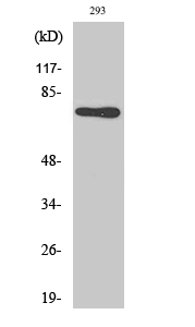
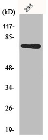
WB analysis of 293 cell lysate using GTX34076 Midline-1 antibody.


WB analysis of 293 cell lysate using GTX34076 Midline-1 antibody.
Midline-1 antibody
GTX34076
ApplicationsImmunoFluorescence, Western Blot, ImmunoCytoChemistry, ImmunoHistoChemistry, ImmunoHistoChemistry Paraffin
Product group Antibodies
ReactivityHuman
TargetMID1
Overview
- SupplierGeneTex
- Product NameMidline-1 antibody
- Delivery Days Customer9
- Application Supplier NoteWB: 1:500-1:2000. ICC/IF: 1:200-1:1000. IHC-P: 1:100-1:300. *Optimal dilutions/concentrations should be determined by the researcher.Not tested in other applications.
- ApplicationsImmunoFluorescence, Western Blot, ImmunoCytoChemistry, ImmunoHistoChemistry, ImmunoHistoChemistry Paraffin
- CertificationResearch Use Only
- ClonalityPolyclonal
- Concentration1 mg/ml
- ConjugateUnconjugated
- Gene ID4281
- Target nameMID1
- Target descriptionmidline 1
- Target synonymsBBBG1, FXY, GBBB, GBBB1, MIDIN, OGS1, OS, OSX, RNF59, TRIM18, XPRF, ZNFXY, E3 ubiquitin-protein ligase Midline-1, Opitz/BBB syndrome, RING finger protein 59, RING finger protein Midline-1, RING-type E3 ubiquitin transferase Midline-1, midline 1 RING finger protein, putative transcription factor XPRF, tripartite motif protein TRIM18, tripartite motif-containing protein 18, zinc finger on X and Y, mouse, homolog of
- HostRabbit
- IsotypeIgG
- Protein IDO15344
- Protein NameE3 ubiquitin-protein ligase Midline-1
- Scientific DescriptionThe protein encoded by this gene is a member of the tripartite motif (TRIM) family, also known as the RING-B box-coiled coil (RBCC) subgroup of RING finger proteins. The TRIM motif includes three zinc-binding domains, a RING, a B-box type 1 and a B-box type 2, and a coiled-coil region. This protein forms homodimers which associate with microtubules in the cytoplasm. The protein is likely involved in the formation of multiprotein structures acting as anchor points to microtubules. Mutations in this gene have been associated with the X-linked form of Opitz syndrome, which is characterized by midline abnormalities such as cleft lip, laryngeal cleft, heart defects, hypospadias, and agenesis of the corpus callosum. This gene was also the first example of a gene subject to X inactivation in human while escaping it in mouse. Alternative promoter use, alternative splicing and alternative polyadenylation result in multiple transcript variants that have different tissue specificities. [provided by RefSeq, Dec 2016]
- ReactivityHuman
- Storage Instruction-20°C or -80°C,2°C to 8°C
- UNSPSC41116161
Datasheet
Related products
Product group Antibodies
MID1 AntibodyCSB-PA003239
ApplicationsImmunoFluorescence, Western Blot, ELISA, ImmunoHistoChemistry
ReactivityHuman, Mouse, Rat
TargetMID1
- SizePrice

Product group Antibodies
Anti-MID1 Antibody Picoband(r)A01774-1-CARRIER-FREE
ApplicationsFlow Cytometry, ImmunoFluorescence, Western Blot, ELISA, ImmunoCytoChemistry, ImmunoHistoChemistry
ReactivityHuman, Mouse
TargetMID1
- SizePrice
Product group Antibodies
Anti-MID1 AntibodyA15627
ApplicationsImmunoFluorescence, Western Blot, ImmunoCytoChemistry
ReactivityHuman, Mouse
- SizePrice
Product group Antibodies

MID1 AntibodyLS-C346337
ApplicationsImmunoFluorescence, Western Blot, ImmunoHistoChemistry
ReactivityHuman, Mouse, Rat
TargetMID1
- SizePrice
Product group Antibodies
Midline-1 antibodyGTX04955
ApplicationsWestern Blot, ImmunoHistoChemistry, ImmunoHistoChemistry Paraffin
ReactivityHuman
TargetMID1
- SizePrice
Product group Antibodies
RNF59/MID1 Polyclonal AntibodyBS-9380R
ApplicationsImmunoFluorescence, Western Blot, ImmunoHistoChemistry, ImmunoHistoChemistry Paraffin
ReactivityCanine, Chicken, Equine, Human, Mouse, Porcine, Rat, Sheep
TargetMID1
- SizePrice