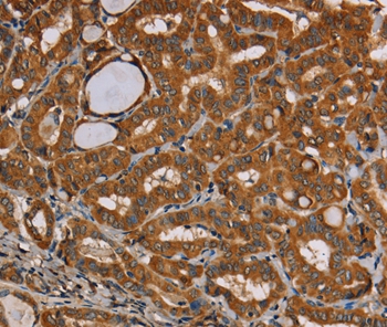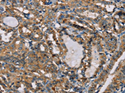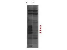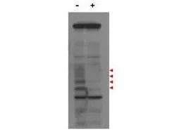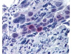
GeneTex's affinity purified anti-MLF1IP pT78 antibody was used at 20 μg/ml to detect signal in a variety of tissues including multi-human, multi-brain and multi-cancer slides. This image shows moderately positive staining of mitotic cells in colon adenocarcinoma at 60X.? Tissue was formalin-fixed and paraffin embedded.? The image shows localization of the antibody as the precipitated red signal, with a hematoxylin purple nuclear counterstain.
Mlf1 Interacting Protein (phospho Thr78) antibody
GTX48727
ApplicationsImmunoFluorescence, Western Blot, ELISA, ImmunoCytoChemistry, ImmunoHistoChemistry, ImmunoHistoChemistry Paraffin
Product group Antibodies
ReactivityHuman
TargetCENPU
Overview
- SupplierGeneTex
- Product NameMlf1 Interacting Protein (phospho Thr78) antibody
- Delivery Days Customer9
- Application Supplier NoteWB: 1:500-1:2000. IHC-P: 20 microg/mL. ELISA: 1:5000-1:25000. *Optimal dilutions/concentrations should be determined by the researcher.Not tested in other applications.
- ApplicationsImmunoFluorescence, Western Blot, ELISA, ImmunoCytoChemistry, ImmunoHistoChemistry, ImmunoHistoChemistry Paraffin
- CertificationResearch Use Only
- ClonalityPolyclonal
- Concentration1 mg/ml
- ConjugateUnconjugated
- Gene ID79682
- Target nameCENPU
- Target descriptioncentromere protein U
- Target synonymsCENP50, CENPU50, KLIP1, MLF1IP, PBIP1, centromere protein U, KSHV latent nuclear antigen interacting protein 1, MLF1 interacting protein, centromere protein of 50 kDa, interphase centromere complex protein 24, polo-box-interacting protein 1
- HostRabbit
- IsotypeIgG
- Protein IDQ71F23
- Protein NameCentromere protein U
- Scientific DescriptionMyeloid leukemia factor-1 (MLF1) Interacting Protein (also known as PBIP1, MLF1IP1, KLIP1 or KSHV latent nuclear antigen interacting protein 1) is a novel polo-like kinase 1 (Plk1) substrate. Plk1 phosphorylation of MLF1IP induces ubiquitination and degradation of MLF1IP prior to the metaphase/ anaphase transition. Several Plk1-dependent phosphorylation sites have been identified on MLF1IP by mass spectrometry. Mutations of these sites stabilize MLF1IP and inhibit mitotic progression. Subsequent in vitro and in vivo MLF1IP phosphorylation and stability assays have revealed that phosphorylation of Thr78 is critical for triggering Plk1-dependent MLF1IP degradation. Expression of a non-degradable Thr78Ala mutant was sufficient to induce a mitotic block. Timely phosphorylation of MLF1IP on Thr78 by Plk1 is critical for eliminating the MLF1IP-imposed mitotic block prior to anaphase onset. MLF1IP is speculated to be a novel tumor suppressor.
- ReactivityHuman
- Storage Instruction-20°C or -80°C,2°C to 8°C
- UNSPSC41116161


