

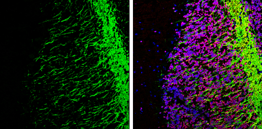
MOG antibody [C2C3], C-term detects MOG protein expression by immunohistochemical analysis. Sample: Frozen-sectioned adult mouse cerebellum. Green: MOG protein stained by MOG antibody [C2C3], C-term (GTX106283) diluted at 1:250. Red: NeuN, stained by NeuN antibody [2Q158] (GTX30773) diluted at 1:500. Blue: Fluoroshield with DAPI (GTX30920).
![MOG antibody [C2C3], C-term detects MOG protein by immunofluorescent analysis. Sample: DIV9 rat E18 primary cortical neurons and glia cells were fixed in 4% paraformaldehyde at RT for 15 min. Green: MOG protein stained by MOG antibody [C2C3], C-term (GTX106283) diluted at 1:500. Red: beta Tubulin 3/ Tuj1, stained by beta Tubulin 3/ Tuj1 antibody [GT886] (GTX631830) diluted at 1:500. Blue: Fluoroshield with DAPI (GTX30920).](https://www.genetex.com/upload/website/prouct_img/normal/GTX106283/GTX106283_39701_20170705_IFA_R_w_23060120_236.webp "MOG antibody [C2C3], C-term detects MOG protein by immunofluorescent analysis. Sample: DIV9 rat E18 primary cortical neurons and glia cells were fixed in 4% paraformaldehyde at RT for 15 min. Green: MOG protein stained by MOG antibody [C2C3], C-term (GTX106283) diluted at 1:500. Red: beta Tubulin 3/ Tuj1, stained by beta Tubulin 3/ Tuj1 antibody [GT886] (GTX631830) diluted at 1:500. Blue: Fluoroshield with DAPI (GTX30920).")
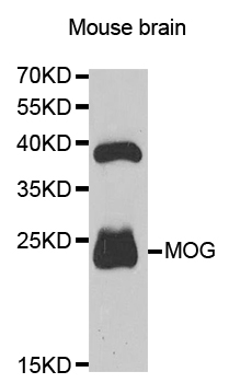
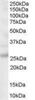
![Various tissue extracts (50 μg) were separated by 12% SDS-PAGE, and the membrane was blotted with MOG antibody [C2C3], C-term (GTX106283) diluted at 1:1000. The HRP-conjugated anti-rabbit IgG antibody (GTX213110-01) was used to detect the primary antibody.](https://www.genetex.com/upload/website/prouct_img/normal/GTX106283/GTX106283_39701_20171006_WB_M_R_w_23060120_130.webp "Various tissue extracts (50 μg) were separated by 12% SDS-PAGE, and the membrane was blotted with MOG antibody [C2C3], C-term (GTX106283) diluted at 1:1000. The HRP-conjugated anti-rabbit IgG antibody (GTX213110-01) was used to detect the primary antibody.")
MOG antibody [C2C3], C-term detects MOG protein expression by immunohistochemical analysis. Sample: Frozen-sectioned adult mouse cerebellum. Green: MOG protein stained by MOG antibody [C2C3], C-term (GTX106283) diluted at 1:250. Red: NeuN, stained by NeuN antibody [2Q158] (GTX30773) diluted at 1:500. Blue: Fluoroshield with DAPI (GTX30920).
MOG antibody [C2C3], C-term
GTX106283
ApplicationsImmunoFluorescence, Western Blot, ImmunoCytoChemistry, ImmunoHistoChemistry, ImmunoHistoChemistry Frozen
Product group Antibodies
ReactivityHuman, Mouse, Rat
TargetMOG
Overview
- SupplierGeneTex
- Product NameMOG antibody [C2C3], C-term
- Delivery Days Customer9
- Application Supplier NoteWB: 1:500-1:3000. ICC/IF: 1:100-1:1000. IHC-Fr: 1:100-1:1000. *Optimal dilutions/concentrations should be determined by the researcher.Not tested in other applications.
- ApplicationsImmunoFluorescence, Western Blot, ImmunoCytoChemistry, ImmunoHistoChemistry, ImmunoHistoChemistry Frozen
- CertificationResearch Use Only
- ClonalityPolyclonal
- Concentration1 mg/ml
- ConjugateUnconjugated
- Gene ID4340
- Target nameMOG
- Target descriptionmyelin oligodendrocyte glycoprotein
- Target synonymsBTN6, BTNL11, MOGIG2, NRCLP7, myelin-oligodendrocyte glycoprotein, MOG AluA, MOG AluB, MOG Ig-AluB, MOG alpha-5
- HostRabbit
- IsotypeIgG
- Protein IDQ16653
- Protein NameMyelin-oligodendrocyte glycoprotein
- Scientific DescriptionThe product of this gene is a membrane protein expressed on the oligodendrocyte cell surface and the outermost surface of myelin sheaths. Due to this localization, it is a primary target antigen involved in immune-mediated demyelination. This protein may be involved in completion and maintenance of the myelin sheath and in cell-cell communication. Alternatively spliced transcript variants encoding different isoforms have been identified. [provided by RefSeq]
- ReactivityHuman, Mouse, Rat
- Storage Instruction-20°C or -80°C,2°C to 8°C
- UNSPSC41116161
Datasheet
Related products
Product group Antibodies
Anti-MOG AntibodyA35668
ApplicationsImmunoFluorescence, Western Blot, ImmunoHistoChemistry
ReactivityHuman, Mouse, Rat
- SizePrice

Product group Antibodies
Anti-Myelin oligodendrocyte glycoprotein/MOG Antibody Picoband(r)A03294-CARRIER-FREE
ApplicationsFlow Cytometry, Western Blot, ImmunoHistoChemistry
ReactivityHuman, Mouse, Rat
TargetMOG
- SizePrice
Product group Antibodies
Anti-MOG IgG Antibody130-10907-20
ApplicationsELISA
ReactivityHuman
TargetMOG
- SizePrice

Product group Antibodies
Anti-MOG AntibodyAMAB91066
ApplicationsWestern Blot, ImmunoHistoChemistry
ReactivityHuman, Mouse, Rat
TargetMOG
- SizePrice
Product group Antibodies

MOG AntibodyLS-C834971
ApplicationsWestern Blot, ELISA, ImmunoHistoChemistry
ReactivityHuman, Mouse, Rat
TargetMOG
- SizePrice
Product group Antibodies
MOG Polyclonal AntibodyBS-0426R
ApplicationsFlow Cytometry, ImmunoFluorescence, Western Blot, ELISA, ImmunoCytoChemistry, ImmunoHistoChemistry, ImmunoHistoChemistry Frozen, ImmunoHistoChemistry Paraffin
ReactivityGuinea Pig, Human, Mouse, Porcine, Rat
TargetMOG
- SizePrice
Product group Antibodies

Goat anti-MOGEB06668
ApplicationsWestern Blot, ELISA
ReactivityBovine, Human, Mouse, Porcine, Rat
TargetMOG
- SizePrice
Product group Antibodies
MOG AntibodyCSB-PA619083ESR2HU
ApplicationsELISA, ImmunoHistoChemistry
ReactivityHuman
TargetMOG
- SizePrice
Product group Antibodies
Mog Polyclonal AntibodyCAC10670
ApplicationsELISA, ImmunoHistoChemistry
TargetMOG
- SizePrice
Product group Antibodies
MOG antibody, C-termGTX89606
ApplicationsWestern Blot, ImmunoHistoChemistry, ImmunoHistoChemistry Paraffin
ReactivityHuman
TargetMOG
- SizePrice