

Mouse anti Human Keratin (4,5,6,8,10,13,18)
X1260M
ApplicationsWestern Blot, ImmunoHistoChemistry, ImmunoHistoChemistry Frozen, ImmunoHistoChemistry Paraffin
Product group Antibodies
TargetKRT5
Overview
- SupplierNordic-MUbio
- Product NameMouse anti Human Keratin (4,5,6,8,10,13,18)
- Delivery Days Customer7
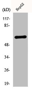
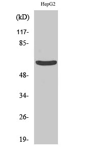
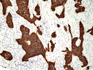
- Application Supplier NoteX1260M detects keratins 4,5,6,8,10,13 & 18 by Western blot. Immunohistochemistry can be performed on frozen tissues and paraffin sections. Optimal concentration should be evaluated by serial dilutions. X1260M reacts with a variety of normal and neoplastic epithelia. Reacting with simple epithelium and both basal and superbasal layers of cornifying and non-cornifying squamous epithelium this antibody is also useful in staining cultured epithelial cell lines. It is useful in differentiating epithelial tumors from non-epithelial tumors.
- ApplicationsWestern Blot, ImmunoHistoChemistry, ImmunoHistoChemistry Frozen, ImmunoHistoChemistry Paraffin
- Applications SupplierImmunohistochemistry (frozen & paraffin);Western Blotting
- CertificationResearch Use Only
- ClonalityMonoclonal
- Clone IDC11
- ConjugateUnconjugated
- Gene ID3852
- Target nameKRT5
- Target descriptionkeratin 5
- Target synonymsCK5, DDD, DDD1, EBS1, EBS2, EBS2A, EBS2B, EBS2C, EBS2D, EBS2E, EBS2F, K5, KRT5A, keratin, type II cytoskeletal 5, 58 kda cytokeratin, CK-5, cytokeratin-5, epidermolysis bullosa simplex 2 Dowling-Meara/Kobner/Weber-Cockayne types, keratin 5 (epidermolysis bullosa simplex, Dowling-Meara/Kobner/Weber-Cockayne types), keratin 5, type II, type-II keratin Kb5
- HostMouse
- IsotypeIgG1
- Protein IDP13647
- Protein NameKeratin, type II cytoskeletal 5
- Shelf life instructionSee expiration date on vial
- SourceHybridoma produced by the fusion of splenocytes from mice immunized with cytoskeleton preparation from human A431 carcinoma cells and mouse myeloma cells.
- Reactivity SupplierHuman
- UNSPSC12352203
Related products
Product group Antibodies
References
Cytokeratin 5 antibodyGTX113219
ApplicationsImmunoFluorescence, Western Blot, ImmunoCytoChemistry, ImmunoHistoChemistry, ImmunoHistoChemistry Paraffin
TargetKRT5
- SizePrice
Product group Antibodies
KRT5 AntibodyCSB-PA002049
ApplicationsImmunoFluorescence, Western Blot, ELISA, ImmunoHistoChemistry
ReactivityHuman, Mouse, Rat
TargetKRT5
- SizePrice
Product group Antibodies

Anti-Basal cell Cytokeratin [RCK103]AB03334-1.1
ApplicationsFlow Cytometry, ImmunoHistoChemistry
TargetKRT5
- SizePrice

Product group Antibodies
Anti-Cytokeratin 5/KRT5 Antibody Picoband(r)A00398-CARRIER-FREE
ApplicationsWestern Blot, ImmunoHistoChemistry
TargetKRT5
- SizePrice
Product group Antibodies
ApplicationsImmunoFluorescence, Western Blot, ELISA, ImmunoHistoChemistry
- SizePrice
Product group Antibodies
Anti-KRT5 Antibody144-02662
ApplicationsImmunoFluorescence, Western Blot, ImmunoHistoChemistry
TargetKRT5
- SizePrice
Product group Antibodies
anti-Cytokeratin-5 (human), Rabbit Monoclonal (RM226)REV-31-1105-00
ApplicationsWestern Blot, ImmunoHistoChemistry
TargetKRT5
- SizePrice

Product group Antibodies
Anti-KRT5 AntibodyAMAB91549
ApplicationsWestern Blot, ImmunoCytoChemistry, ImmunoHistoChemistry
ReactivityHuman
TargetKRT5
- SizePrice