

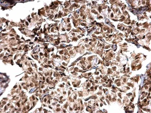
MTA1 antibody [C1C3] detects MTA1 protein at nucleus on human breast carcinoma by immunohistochemical analysis. Sample: Paraffin-embedded human breast carcinoma. MTA1 antibody [C1C3] (GTX100610) dilution: 1:500.
Antigen Retrieval: Trilogy? (EDTA based, pH 8.0) buffer, 15min
![MTA1 antibody [C1C3] detects MTA1 protein at nucleus by immunofluorescent analysis. Sample: A431 cells were fixed in 4% paraformaldehyde at RT for 15 min. Green: MTA1 protein stained by MTA1 antibody [C1C3] (GTX100610) diluted at 1:500. Blue: Hoechst 33342 staining.](https://www.genetex.com/upload/website/prouct_img/normal/GTX100610/GTX100610_41073_IFA_w_23060100_389.webp "MTA1 antibody [C1C3] detects MTA1 protein at nucleus by immunofluorescent analysis. Sample: A431 cells were fixed in 4% paraformaldehyde at RT for 15 min. Green: MTA1 protein stained by MTA1 antibody [C1C3] (GTX100610) diluted at 1:500. Blue: Hoechst 33342 staining.")
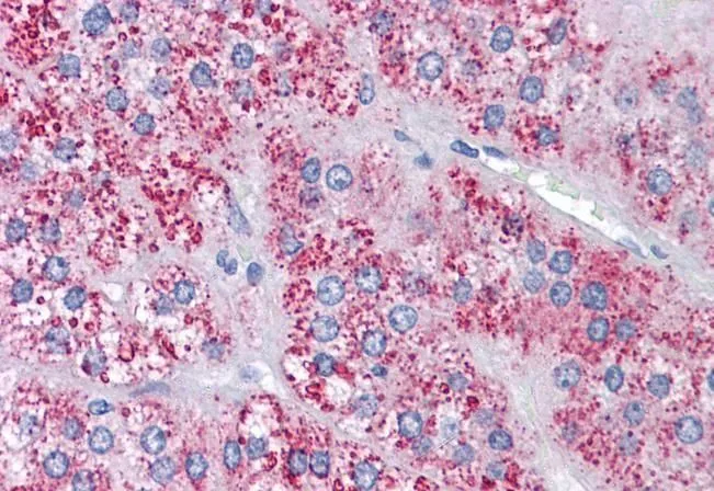
![MTA1 antibody [C1C3] detects MTA1 protein at nucleus on mouse ovary by immunohistochemical analysis. Sample: Paraffin-embedded mouse ovary. MTA1 antibody [C1C3] (GTX100610) dilution: 1:500.
Antigen Retrieval: Trilogy? (EDTA based, pH 8.0) buffer, 15min](https://www.genetex.com/upload/website/prouct_img/normal/GTX100610/GTX100610_41073_IHC_M_w_23060100_714.webp "MTA1 antibody [C1C3] detects MTA1 protein at nucleus on mouse ovary by immunohistochemical analysis. Sample: Paraffin-embedded mouse ovary. MTA1 antibody [C1C3] (GTX100610) dilution: 1:500.
Antigen Retrieval: Trilogy? (EDTA based, pH 8.0) buffer, 15min")
![MTA1 antibody [C1C3] detects MTA1 protein at nucleus on rat hind brain by immunohistochemical analysis. Sample: Paraffin-embedded rat hind brain. MTA1 antibody [C1C3] (GTX100610) dilution: 1:500.
Antigen Retrieval: Trilogy? (EDTA based, pH 8.0) buffer, 15min](https://www.genetex.com/upload/website/prouct_img/normal/GTX100610/GTX100610_41073_IHC_R_w_23060100_884.webp "MTA1 antibody [C1C3] detects MTA1 protein at nucleus on rat hind brain by immunohistochemical analysis. Sample: Paraffin-embedded rat hind brain. MTA1 antibody [C1C3] (GTX100610) dilution: 1:500.
Antigen Retrieval: Trilogy? (EDTA based, pH 8.0) buffer, 15min")
MTA1 antibody [C1C3] detects MTA1 protein at nucleus on human breast carcinoma by immunohistochemical analysis. Sample: Paraffin-embedded human breast carcinoma. MTA1 antibody [C1C3] (GTX100610) dilution: 1:500.
Antigen Retrieval: Trilogy? (EDTA based, pH 8.0) buffer, 15min
MTA1 antibody [C1C3]
GTX100610
ApplicationsImmunoFluorescence, ImmunoCytoChemistry, ImmunoHistoChemistry, ImmunoHistoChemistry Paraffin
Product group Antibodies
ReactivityHuman, Mouse, Rat
TargetMTA1
Overview
- SupplierGeneTex
- Product NameMTA1 antibody [C1C3]
- Delivery Days Customer9
- Application Supplier NoteICC/IF: 1:100-1:1000. IHC-P: 1:100-1:1000. *Optimal dilutions/concentrations should be determined by the researcher.Not tested in other applications.
- ApplicationsImmunoFluorescence, ImmunoCytoChemistry, ImmunoHistoChemistry, ImmunoHistoChemistry Paraffin
- CertificationResearch Use Only
- ClonalityPolyclonal
- Concentration1 mg/ml
- ConjugateUnconjugated
- Gene ID9112
- Target nameMTA1
- Target descriptionmetastasis associated 1
- Target synonymsmetastasis-associated protein MTA1, metastasis associated gene 1 protein
- HostRabbit
- IsotypeIgG
- Protein IDQ13330
- Protein NameMetastasis-associated protein MTA1
- Scientific DescriptionThis gene encodes a protein that was identified in a screen for genes expressed in metastatic cells, specifically, mammary adenocarcinoma cell lines. Expression of this gene has been correlated with the metastatic potential of at least two types of carcinomas although it is also expressed in many normal tissues. The role it plays in metastasis is unclear. It was initially thought to be the 70kD component of a nucleosome remodeling deacetylase complex, NuRD, but it is more likely that this component is a different but very similar protein. These two proteins are so closely related, though, that they share the same types of domains. These domains include two DNA binding domains, a dimerization domain, and a domain commonly found in proteins that methylate DNA. The profile and activity of this gene product suggest that it is involved in regulating transcription and that this may be accomplished by chromatin remodeling. [provided by RefSeq]
- ReactivityHuman, Mouse, Rat
- Storage Instruction-20°C or -80°C,2°C to 8°C
- UNSPSC41116161
Datasheet
Related products
Product group Antibodies
Anti-MTA1 AntibodyA98029
ApplicationsWestern Blot, ELISA
ReactivityHuman, Mouse, Rat
- SizePrice
Product group Antibodies

Anti-MTA1 [RAB-T21]Ab01812-1.1
ApplicationsFlow Cytometry, ImmunoFluorescence, ImmunoPrecipitation
ReactivityHuman
TargetMTA1
- SizePrice
Product group Antibodies
Anti-MTA1 Antibody144-64083
ApplicationsWestern Blot
ReactivityHuman, Mouse
TargetMTA1
- SizePrice
Product group Antibodies

Goat anti-MTA1EB05163
ApplicationsWestern Blot, ELISA, ImmunoHistoChemistry
ReactivityHuman
TargetMTA1
- SizePrice
Product group Antibodies
MTA1 AntibodyCSB-PA070230
ApplicationsWestern Blot, ELISA, ImmunoHistoChemistry
ReactivityHuman, Mouse, Rat
TargetMTA1
- SizePrice
Product group Antibodies
MTA1 Polyclonal AntibodyBS-1412R
ApplicationsImmunoFluorescence, Western Blot, ELISA, ImmunoCytoChemistry, ImmunoHistoChemistry, ImmunoHistoChemistry Frozen, ImmunoHistoChemistry Paraffin
ReactivityHuman, Mouse, Rat
TargetMTA1
- SizePrice
Product group Antibodies
MTA1 antibody, InternalGTX20751
ApplicationsWestern Blot, ImmunoHistoChemistry, ImmunoHistoChemistry Paraffin
ReactivityHuman
TargetMTA1
- SizePrice
Product group Antibodies

MTA1 Antibody (Internal)LS-C358813
ApplicationsWestern Blot, ImmunoHistoChemistry, ImmunoHistoChemistry Paraffin
ReactivityHuman, Mouse, Rat
TargetMTA1
- SizePrice

Product group Antibodies
Anti-MTA1 AntibodyHPA005544
ApplicationsImmunoCytoChemistry, ImmunoHistoChemistry
ReactivityHuman
TargetMTA1
- SizePrice
Product group Antibodies
MTA1 antibodyGTX66656
ApplicationsWestern Blot
ReactivityHuman, Mouse, Rat
TargetMTA1
- SizePrice