

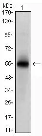
WB analysis of human MSI1 (AA: 1-203) recombinant protein using GTX60408 Musashi 1 antibody [2A12].
![FACS analysis of PC-2 cells using GTX60408 Musashi 1 antibody [2A12]. Green : Musashi 1 Purple : negative control](https://www.genetex.com/upload/website/prouct_img/normal/GTX60408/GTX60408_20170912_FACS_w_23061123_121.webp "FACS analysis of PC-2 cells using GTX60408 Musashi 1 antibody [2A12]. Green : Musashi 1 Purple : negative control")
![ELISA analysis of antigen using GTX60408 Musashi 1 antibody [2A12].
Red : Control antigen 100ng
Purple : Antigen 10ng
Green : Antigen 50ng
Blue : Antigen 100ng](https://www.genetex.com/upload/website/prouct_img/normal/GTX60408/GTX60408_20170912_ELISA_w_23061123_511.webp "ELISA analysis of antigen using GTX60408 Musashi 1 antibody [2A12].
Red : Control antigen 100ng
Purple : Antigen 10ng
Green : Antigen 50ng
Blue : Antigen 100ng")
![WB analysis of NTERA-2 cell lysate using GTX60408 Musashi 1 antibody [2A12].](https://www.genetex.com/upload/website/prouct_img/normal/GTX60408/GTX60408_20170912_WB_1_w_23061123_854.webp "WB analysis of NTERA-2 cell lysate using GTX60408 Musashi 1 antibody [2A12].")
WB analysis of human MSI1 (AA: 1-203) recombinant protein using GTX60408 Musashi 1 antibody [2A12].
Musashi 1 antibody [2A12]
GTX60408
ApplicationsFlow Cytometry, Western Blot, ELISA
Product group Antibodies
ReactivityHuman
TargetMSI1
Overview
- SupplierGeneTex
- Product NameMusashi 1 antibody [2A12]
- Delivery Days Customer9
- Application Supplier NoteWB: 1/500 - 1/2000. FCM: 1/200 - 1/400. ELISA: 1/10000. *Optimal dilutions/concentrations should be determined by the researcher.Not tested in other applications.
- ApplicationsFlow Cytometry, Western Blot, ELISA
- CertificationResearch Use Only
- ClonalityMonoclonal
- Clone ID2A12
- ConjugateUnconjugated
- Gene ID4440
- Target nameMSI1
- Target descriptionmusashi RNA binding protein 1
- Target synonymsRNA-binding protein Musashi homolog 1, musashi-1, musashi1
- HostMouse
- IsotypeIgG1
- Protein IDO43347
- Protein NameRNA-binding protein Musashi homolog 1
- Scientific DescriptionThis gene encodes a protein containing two conserved tandem RNA recognition motifs. Similar proteins in other species function as RNA-binding proteins and play central roles in posttranscriptional gene regulation. Expression of this gene has been correlated with the grade of the malignancy and proliferative activity in gliomas and melanomas. A pseudogene for this gene is located on chromosome 11q13. [provided by RefSeq, Jul 2008]
- ReactivityHuman
- Storage Instruction-20°C or -80°C,2°C to 8°C
- UNSPSC41116161
Datasheet
Related products
Product group Antibodies
Anti-Musashi 1/Msi1 Antibody Picoband(r)A05052-1-CARRIER-FREE
ApplicationsFlow Cytometry, ImmunoFluorescence, Western Blot, ImmunoCytoChemistry, ImmunoHistoChemistry
ReactivityHuman, Mouse, Rat
TargetMSI1
- SizePrice
Product group Antibodies
Anti-MSI1 Antibody144-65558
ApplicationsImmunoFluorescence, Western Blot, ImmunoHistoChemistry
ReactivityHuman, Mouse, Rat
TargetMSI1
- SizePrice
Product group Antibodies
Anti-Musashi-1 AntibodyA32522
ApplicationsWestern Blot
ReactivityHuman, Mouse, Rat
- SizePrice
Product group Antibodies

MSI1 / Musashi 1 AntibodyLS-C768281
ApplicationsWestern Blot, ELISA, ImmunoHistoChemistry
ReactivityHuman, Mouse, Rat
TargetMSI1
- SizePrice
Product group Antibodies
Musashi 1 Polyclonal AntibodyBS-11201R
ApplicationsFlow Cytometry, ImmunoFluorescence, ImmunoCytoChemistry, ImmunoHistoChemistry, ImmunoHistoChemistry Frozen, ImmunoHistoChemistry Paraffin
TargetMSI1
- SizePrice
Product group Antibodies
MSI1 AntibodyCSB-PA015033LA01HU
ApplicationsImmunoFluorescence, ELISA, ImmunoHistoChemistry
ReactivityHuman
TargetMSI1
- SizePrice
![MSI1 antibody [N3C3] detects MSI1 protein at nucleus by immunofluorescent analysis. Sample: HeLa cells were fixed in 4% paraformaldehyde at RT for 15 min. Green: MSI1 protein stained by MSI1 antibody [N3C3] (GTX101540) diluted at 1:500. Red: alpha Tubulin, a cytoskeleton marker, stained by alpha Tubulin antibody [GT114] (GTX628802) diluted at 1:1000.](https://www.genetex.com/upload/website/prouct_img/normal/GTX101540/GTX101540_39911_20150410_IFA_w_23060100_981.webp)
Product group Antibodies
Musashi 1 antibody [N3C3]GTX101540
ApplicationsImmunoFluorescence, ImmunoPrecipitation, Western Blot, ImmunoCytoChemistry, ImmunoHistoChemistry, ImmunoHistoChemistry Frozen
ReactivityHuman, Mouse
TargetMSI1
- SizePrice
Product group Antibodies
Musashi 1 antibodyGTX78273
ApplicationsImmunoFluorescence, Western Blot, ImmunoCytoChemistry, ImmunoHistoChemistry, ImmunoHistoChemistry Paraffin
ReactivityHuman, Mouse, Rat
- SizePrice

Product group Antibodies
Anti-MSI1 AntibodyHPA064401
ApplicationsImmunoCytoChemistry
ReactivityHuman
TargetMSI1
- SizePrice
![Mouse tissue extract (50 μg) was separated by 12% SDS-PAGE, and the membrane was blotted with Musashi 1 antibody [GT2377] (GTX634485) diluted at 1:1000. The HRP-conjugated anti-mouse IgG antibody (GTX213111-01) was used to detect the primary antibody.](https://www.genetex.com/upload/website/prouct_img/normal/GTX634485/GTX634485_43257_20180713_WB_M_fetalbrain_w_23061202_875.webp)
Product group Antibodies
Musashi 1 antibody [GT2377]GTX634485
ApplicationsWestern Blot
ReactivityHuman, Mouse
TargetMSI1
- SizePrice