

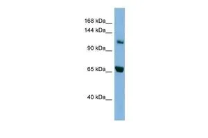
WB analysis of HepG2 cells using GTX45049 MYO1E antibody at 0.2-1μg/ml.
WB analysis of HepG2 cells using GTX45049 MYO1E antibody at 0.2-1μg/ml.
MYO1E antibody, Internal
GTX45049
ApplicationsWestern Blot
Product group Antibodies
ReactivityHuman
TargetMYO1E
Overview
- SupplierGeneTex
- Product NameMYO1E antibody, Internal
- Delivery Days Customer9
- Application Supplier NoteWB: 0.2-2.5 ug/ml. *Optimal dilutions/concentrations should be determined by the researcher.Not tested in other applications.
- ApplicationsWestern Blot
- CertificationResearch Use Only
- ClonalityPolyclonal
- Concentration0.5-1 mg/ml
- ConjugateUnconjugated
- Gene ID4643
- Target nameMYO1E
- Target descriptionmyosin IE
- Target synonymsFSGS6, HuncM-IC, MYO1C, unconventional myosin-Ie, MYO1E variant protein, myosin-IC, unconventional myosin 1E
- HostRabbit
- IsotypeIgG
- Protein IDQ12965
- Protein NameUnconventional myosin-Ie
- Scientific DescriptionThis gene encodes a member of the nonmuscle class I myosins which are a subgroup of the unconventional myosin protein family. The unconventional myosin proteins function as actin-based molecular motors. Class I myosins are characterized by a head (motor) domain, a regulatory domain and a either a short or long tail domain. Among the class I myosins, this protein is distinguished by a long tail domain that is involved in crosslinking actin filaments. This protein localizes to the cytoplasm and may be involved in intracellular movement and membrane trafficking. Mutations in this gene are the cause of focal segmental glomerulosclerosis-6. This gene has been referred to as myosin IC in the literature but is distinct from the myosin IC gene located on chromosome 17. [provided by RefSeq, Jan 2012]
- ReactivityHuman
- Storage Instruction-20°C or -80°C,2°C to 8°C
- UNSPSC41116161
Datasheet
Related products
Product group Antibodies

MYO1E / Myosin IE AntibodyLS-C748095
ApplicationsWestern Blot
ReactivityHuman
TargetMYO1E
- SizePrice
Product group Antibodies
MYO1E AntibodyCSB-PA621526LA01HU
ApplicationsELISA, ImmunoHistoChemistry
ReactivityHuman
TargetMYO1E
- SizePrice
Product group Antibodies
ApplicationsImmunoPrecipitation, Western Blot, ImmunoCytoChemistry, ImmunoHistoChemistry
ReactivityMouse, Rat
TargetMYO1E
- SizePrice

Product group Antibodies
Anti-MYO1E AntibodyHPA023886
ApplicationsWestern Blot, ImmunoHistoChemistry
ReactivityHuman
TargetMYO1E
- SizePrice