

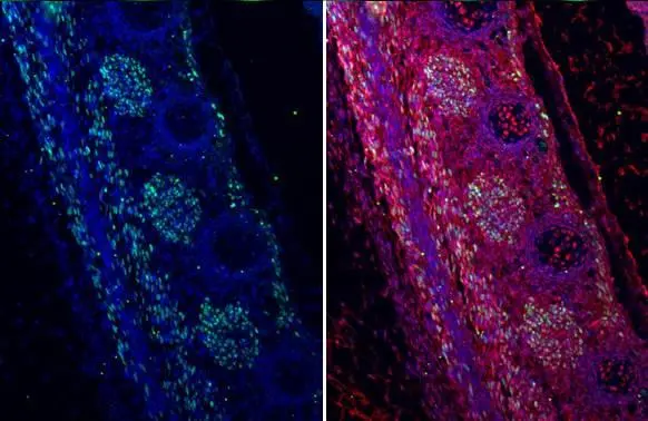
MyoD1 antibody [HL1372] detects MyoD1 protein at nucleus by immunohistochemical analysis. Sample: Paraffin-embedded mouse E13.5 embryo. Green: MyoD1 stained by MyoD1 antibody [HL1372] (GTX636812) diluted at 1:100. Red: SOX2, a nucleus marker, stained by SOX2 antibody [GT1352] (GTX627405) diluted at 1:250. Blue: Fluoroshield with DAPI (GTX30920). Antigen Retrieval: Citrate buffer, pH 6.0, 15 min
![MyoD1 antibody [HL1372] detects MyoD1 protein at nucleus by immunofluorescent analysis. Sample: RMS-13 cells were fixed in 4% paraformaldehyde at RT for 15 min. Green: MyoD1 stained by MyoD1 antibody [HL1372] (GTX636812) diluted at 1:500. Red: alpha Tubulin, a cytoskeleton marker, stained by alpha Tubulin antibody [GT114] (GTX628802) diluted at 1:1000.](https://www.genetex.com/upload/website/prouct_img/normal/GTX636812/GTX636812_44669_20220701_ICC_IF_22071401_453.webp "MyoD1 antibody [HL1372] detects MyoD1 protein at nucleus by immunofluorescent analysis. Sample: RMS-13 cells were fixed in 4% paraformaldehyde at RT for 15 min. Green: MyoD1 stained by MyoD1 antibody [HL1372] (GTX636812) diluted at 1:500. Red: alpha Tubulin, a cytoskeleton marker, stained by alpha Tubulin antibody [GT114] (GTX628802) diluted at 1:1000.")
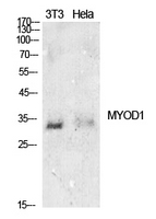
![Various tissue extracts (30 μg) were separated by 10% SDS-PAGE, and the membrane was blotted with MyoD1 antibody [HL1372] (GTX636812) diluted at 1:1000. The HRP-conjugated anti-rabbit IgG antibody (GTX213110-01) was used to detect the primary antibody.](https://www.genetex.com/upload/website/prouct_img/normal/GTX636812/GTX636812_T-44606_20220325_WB_M_R_w_23061202_291.webp "Various tissue extracts (30 μg) were separated by 10% SDS-PAGE, and the membrane was blotted with MyoD1 antibody [HL1372] (GTX636812) diluted at 1:1000. The HRP-conjugated anti-rabbit IgG antibody (GTX213110-01) was used to detect the primary antibody.")
![Non-transfected (–) and transfected (+) 293T whole cell extracts (30 μg) were separated by 12% SDS-PAGE, and the membrane was blotted with MyoD1 antibody [HL1372] (GTX636812) diluted at 1:15000. The HRP-conjugated anti-rabbit IgG antibody (GTX213110-01) was used to detect the primary antibody.](https://www.genetex.com/upload/website/prouct_img/normal/GTX636812/GTX636812_44669_20231215_WB_multiple_B_23121922_430.webp "Non-transfected (–) and transfected (+) 293T whole cell extracts (30 μg) were separated by 12% SDS-PAGE, and the membrane was blotted with MyoD1 antibody [HL1372] (GTX636812) diluted at 1:15000. The HRP-conjugated anti-rabbit IgG antibody (GTX213110-01) was used to detect the primary antibody.")
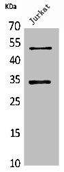
![Various whole cell extracts (30 μg) were separated by 10% SDS-PAGE, and the membrane was blotted with MyoD1 antibody [HL1372] (GTX636812) diluted at 1:2000. The HRP-conjugated anti-rabbit IgG antibody (GTX213110-01) was used to detect the primary antibody. Corresponding RNA expression data for the same cell lines are based on Human Protein Atlas program.](https://www.genetex.com/upload/website/prouct_img/normal/GTX636812/GTX636812_45474_20240719_WB_TPM_watermark_25071701_223.webp "Various whole cell extracts (30 μg) were separated by 10% SDS-PAGE, and the membrane was blotted with MyoD1 antibody [HL1372] (GTX636812) diluted at 1:2000. The HRP-conjugated anti-rabbit IgG antibody (GTX213110-01) was used to detect the primary antibody. Corresponding RNA expression data for the same cell lines are based on Human Protein Atlas program.")
MyoD1 antibody [HL1372] detects MyoD1 protein at nucleus by immunohistochemical analysis. Sample: Paraffin-embedded mouse E13.5 embryo. Green: MyoD1 stained by MyoD1 antibody [HL1372] (GTX636812) diluted at 1:100. Red: SOX2, a nucleus marker, stained by SOX2 antibody [GT1352] (GTX627405) diluted at 1:250. Blue: Fluoroshield with DAPI (GTX30920). Antigen Retrieval: Citrate buffer, pH 6.0, 15 min
MyoD1 antibody [HL1372]
GTX636812
ApplicationsImmunoFluorescence, Western Blot, ImmunoCytoChemistry, ImmunoHistoChemistry, ImmunoHistoChemistry Paraffin
Product group Antibodies
ReactivityHuman, Mouse, Rat
TargetMYOD1
Overview
- SupplierGeneTex
- Product NameMyoD1 antibody [HL1372]
- Delivery Days Customer9
- Application Supplier NoteWB: 1:500-1:3000. *Optimal dilutions/concentrations should be determined by the researcher.Not tested in other applications.
- ApplicationsImmunoFluorescence, Western Blot, ImmunoCytoChemistry, ImmunoHistoChemistry, ImmunoHistoChemistry Paraffin
- CertificationResearch Use Only
- ClonalityMonoclonal
- Clone IDHL1372
- Concentration1 mg/ml
- ConjugateUnconjugated
- Gene ID4654
- Target nameMYOD1
- Target descriptionmyogenic differentiation 1
- Target synonymsCMYO17, CMYP17, MYF3, MYOD, MYODRIF, PUM, bHLHc1, myoblast determination protein 1, class C basic helix-loop-helix protein 1, myf-3, myogenic factor 3
- HostRabbit
- IsotypeIgG
- Protein IDP15172
- Protein NameMyoblast determination protein 1
- Scientific DescriptionThis gene encodes a nuclear protein that belongs to the basic helix-loop-helix family of transcription factors and the myogenic factors subfamily. It regulates muscle cell differentiation by inducing cell cycle arrest, a prerequisite for myogenic initiation. The protein is also involved in muscle regeneration. It activates its own transcription which may stabilize commitment to myogenesis. [provided by RefSeq, Jul 2008]
- ReactivityHuman, Mouse, Rat
- Storage Instruction-20°C or -80°C,2°C to 8°C
- UNSPSC41116161
Datasheet
Related products
Product group Antibodies
Anti-MYOD1 AntibodyA97396
ApplicationsWestern Blot, ELISA
ReactivityHuman, Mouse, Rat
- SizePrice
Product group Antibodies

Anti-MYOD1 [6F4-2D7]Ab03307-10.0
ApplicationsWestern Blot, ELISA, ImmunoHistoChemistry
ReactivityHuman
TargetMYOD1
- SizePrice
Product group Antibodies
Anti-MyoD1 Antibody188-11549
ApplicationsFlow Cytometry
ReactivityChicken, Human, Mouse, Rat
TargetMYOD1
- SizePrice
Product group Antibodies

MYOD / MYOD1 Antibody (clone ABT-MYOD1)LS-C744545
ApplicationsWestern Blot, ImmunoHistoChemistry
ReactivityHuman
TargetMYOD1
- SizePrice
Product group Antibodies
MyoD1/Myf3 Polyclonal AntibodyBS-23809R
ApplicationsWestern Blot, ImmunoHistoChemistry, ImmunoHistoChemistry Paraffin
ReactivityBovine, Canine, Equine, Human, Mouse, Porcine, Rat, Sheep
TargetMYOD1
- SizePrice
Product group Antibodies
MYOD1 Polyclonal AntibodyCAC14646
ApplicationsWestern Blot, ELISA
ReactivityMouse
TargetMYOD1
- SizePrice
Product group Antibodies
MYOD1 AntibodyCSB-PA005516
ApplicationsWestern Blot, ELISA
ReactivityHuman, Mouse, Rat
TargetMYOD1
- SizePrice

Product group Antibodies
ApplicationsFlow Cytometry, ImmunoPrecipitation, Western Blot
ReactivityHuman
TargetMYOD1
- SizePrice
![IHC-P analysis of human rhabdomyosarcoma tissue using GTX17712 MyoD1 antibody [MYOD1/2075R].](https://www.genetex.com/upload/website/prouct_img/normal/GTX17712/GTX17712_20200115_IHC-P_1089_w_23060620_737.webp)
Product group Antibodies
MyoD1 antibody [MYOD1/2075R]GTX17712
ApplicationsImmunoHistoChemistry, ImmunoHistoChemistry Paraffin, Other Application
ReactivityHuman
TargetMYOD1
- SizePrice
![IHC-P analysis of human rhabdomyosarcoma tissue using GTX17751 MyoD1 antibody [rMYD712].](https://www.genetex.com/upload/website/prouct_img/normal/GTX17751/GTX17751_20200115_IHC-P_1091_w_23060620_304.webp)
Product group Antibodies
MyoD1 antibody [rMYD712]GTX17751
ApplicationsImmunoHistoChemistry, ImmunoHistoChemistry Paraffin, Other Application
ReactivityHuman
TargetMYOD1
- SizePrice