

WB analysis of various samples using GTX03240 N-WASP antibody [GT1328]. Dilution : 1:1000 Loading : 25μg per lane
![IHC-P analysis of mouse kidney tissue section using GTX03240 N-WASP antibody [GT1328]. Dilution : 1:100](https://www.genetex.com/upload/website/prouct_img/normal/GTX03240/GTX03240_20210615_IHC-P_17_w_23053123_539.webp "IHC-P analysis of mouse kidney tissue section using GTX03240 N-WASP antibody [GT1328]. Dilution : 1:100")
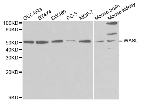
![Various whole cell extracts (30 μg) were separated by 7.5% SDS-PAGE, and the membrane was blotted with N-WASP antibody [GT1328] (GTX03240) diluted at 1:1000. The HRP-conjugated anti-rabbit IgG antibody (GTX213110-01) was used to detect the primary antibody.](https://www.genetex.com/upload/website/prouct_img/normal/GTX03240/GTX03240_4000000737_20210716_WB_2_w_23053123_603.webp "Various whole cell extracts (30 μg) were separated by 7.5% SDS-PAGE, and the membrane was blotted with N-WASP antibody [GT1328] (GTX03240) diluted at 1:1000. The HRP-conjugated anti-rabbit IgG antibody (GTX213110-01) was used to detect the primary antibody.")
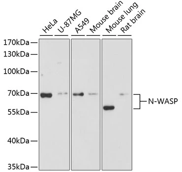
![WB analysis of HeLa whole cell lysate using GTX03240 N-WASP antibody [GT1328]. Dilution : 1:1000 Loading : 25μg per lane](https://www.genetex.com/upload/website/prouct_img/normal/GTX03240/GTX03240_21_WB_w_23053123_619.webp "WB analysis of HeLa whole cell lysate using GTX03240 N-WASP antibody [GT1328]. Dilution : 1:1000 Loading : 25μg per lane")
![IHC-P analysis of human placenta tissue section using GTX03240 N-WASP antibody [GT1328]. Dilution : 1:100](https://www.genetex.com/upload/website/prouct_img/normal/GTX03240/GTX03240_20210615_IHC-P_16_w_23053123_824.webp "IHC-P analysis of human placenta tissue section using GTX03240 N-WASP antibody [GT1328]. Dilution : 1:100")
![Various whole cell extracts (30 μg) were separated by 7.5% SDS-PAGE, and the membrane was blotted with N-WASP antibody [GT1328] (GTX03240) diluted at 1:1000. The HRP-conjugated anti-rabbit IgG antibody (GTX213110-01) was used to detect the primary antibody.](https://www.genetex.com/upload/website/prouct_img/normal/GTX03240/GTX03240_4000000737_20210716_WB_w_23053123_764.webp "Various whole cell extracts (30 μg) were separated by 7.5% SDS-PAGE, and the membrane was blotted with N-WASP antibody [GT1328] (GTX03240) diluted at 1:1000. The HRP-conjugated anti-rabbit IgG antibody (GTX213110-01) was used to detect the primary antibody.")
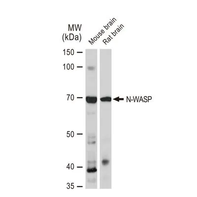
WB analysis of various samples using GTX03240 N-WASP antibody [GT1328]. Dilution : 1:1000 Loading : 25μg per lane
N-WASP antibody [GT1328]
GTX03240
ApplicationsWestern Blot, ImmunoHistoChemistry, ImmunoHistoChemistry Paraffin
Product group Antibodies
ReactivityHuman, Mouse, Rat
TargetWASL
Overview
- SupplierGeneTex
- Product NameN-WASP antibody [GT1328]
- Delivery Days Customer9
- Application Supplier NoteWB: 1:500 - 1:2000. IHC-P: 1:50 - 1:200. *Optimal dilutions/concentrations should be determined by the researcher.Not tested in other applications.
- ApplicationsWestern Blot, ImmunoHistoChemistry, ImmunoHistoChemistry Paraffin
- CertificationResearch Use Only
- ClonalityMonoclonal
- Clone IDGT1328
- Concentration1.8 mg/ml
- ConjugateUnconjugated
- Gene ID8976
- Target nameWASL
- Target descriptionWASP like actin nucleation promoting factor
- Target synonymsN-WASP, NWASP, WASPB, actin nucleation-promoting factor WASL, Wiskott-Aldrich syndrome like, neural Wiskott-Aldrich syndrome protein
- HostRabbit
- IsotypeIgG
- Protein IDO00401
- Protein NameActin nucleation-promoting factor WASL
- Scientific DescriptionThis gene encodes a member of the Wiskott-Aldrich syndrome (WAS) protein family. Wiskott-Aldrich syndrome proteins share similar domain structure, and associate with a variety of signaling molecules to alter the actin cytoskeleton. The encoded protein is highly expressed in neural tissues, and interacts with several proteins involved in cytoskeletal organization, including cell division control protein 42 (CDC42) and the actin-related protein-2/3 (ARP2/3) complex. The encoded protein may be involved in the formation of long actin microspikes, and in neurite extension. [provided by RefSeq, Jul 2013]
- ReactivityHuman, Mouse, Rat
- Storage Instruction-20°C or -80°C,2°C to 8°C
- UNSPSC41116161
Datasheet
Related products
Product group Antibodies
WASL AntibodyCSB-PA025973GA01HU
ApplicationsImmunoFluorescence, Western Blot, ELISA, ImmunoHistoChemistry
ReactivityHuman, Mouse, Rat
TargetWASL
- SizePrice
Product group Antibodies
Anti-WASL Antibody144-02576
ApplicationsWestern Blot
ReactivityHuman, Mouse, Rat
TargetWASL
- SizePrice

Product group Antibodies
Anti-N WASP/WASL Antibody Picoband(r)A05438-3-CARRIER-FREE
ApplicationsFlow Cytometry, Western Blot, ELISA
ReactivityHuman, Mouse, Rat
TargetWASL
- SizePrice
Product group Antibodies
Anti-WASL AntibodyA30588
ApplicationsImmunoFluorescence, Western Blot, ImmunoHistoChemistry
ReactivityHuman, Mouse, Rat
- SizePrice

Product group Antibodies
Anti-WASL AntibodyHPA005750
ApplicationsWestern Blot, ImmunoCytoChemistry, ImmunoHistoChemistry
ReactivityHuman, Mouse, Rat
TargetWASL
- SizePrice
Product group Antibodies

WASL / N-WASP AntibodyLS-C332179
ApplicationsImmunoFluorescence, ImmunoPrecipitation, Western Blot, ImmunoHistoChemistry
ReactivityHuman, Mouse, Rat
TargetWASL
- SizePrice
Product group Antibodies
N-WASP antibodyGTX32762
ApplicationsImmunoFluorescence, Western Blot, ImmunoCytoChemistry, ImmunoHistoChemistry, ImmunoHistoChemistry Paraffin
ReactivityHuman, Mouse, Rat
TargetWASL
- SizePrice
Product group Antibodies
N WASP Recombinant Antibody, Biotin ConjugatedBSM-61522R-BIOTIN
ApplicationsWestern Blot, ImmunoHistoChemistry, ImmunoHistoChemistry Frozen, ImmunoHistoChemistry Paraffin
ReactivityHuman, Mouse
TargetWASL
- SizePrice