

Sample (30 μg of whole cell lysate) A: 293T B: A431 C: HeLa D: HepG2 10% SDS PAGE GTX627421 diluted at 1:1000 The HRP-conjugated anti-mouse IgG antibody (GTX213111-01) was used to detect the primary antibody.
![NANOG antibody [GT3312] validation by siRNA knock-down. Upperpanel: NANOG antibody [GT3312] GTX627421 Lower panel: GAPDH antibody (GTX100118) A. 30 μg Tera-2 whole cell lysate/extract B. 30 μg whole cell lysate/extract of NANOG siRNA#1-transfected Tera-2 cells C. 30 μg whole cell lysate/extract of NANOG siRNA#2-transfected Tera-2 cells D. 30 μg whole cell lysate/extract of NANOG siRNA#3-transfected Tera-2 cells 10% SDS-PAGE NANOG antibody [GT3312] (GTX627421) dilution: 1:1000 GAPDH antibody (GTX100118) dilution: 1:10000 The HRP-conjugated anti-mouse IgG antibody (GTX213111-01) was used to detect the primary antibody.](https://www.genetex.com/upload/website/prouct_img/normal/GTX627421/GTX627421_40878_WB_siRNA_w_23061202_558.webp "NANOG antibody [GT3312] validation by siRNA knock-down. Upperpanel: NANOG antibody [GT3312] GTX627421 Lower panel: GAPDH antibody (GTX100118) A. 30 μg Tera-2 whole cell lysate/extract B. 30 μg whole cell lysate/extract of NANOG siRNA#1-transfected Tera-2 cells C. 30 μg whole cell lysate/extract of NANOG siRNA#2-transfected Tera-2 cells D. 30 μg whole cell lysate/extract of NANOG siRNA#3-transfected Tera-2 cells 10% SDS-PAGE NANOG antibody [GT3312] (GTX627421) dilution: 1:1000 GAPDH antibody (GTX100118) dilution: 1:10000 The HRP-conjugated anti-mouse IgG antibody (GTX213111-01) was used to detect the primary antibody.")
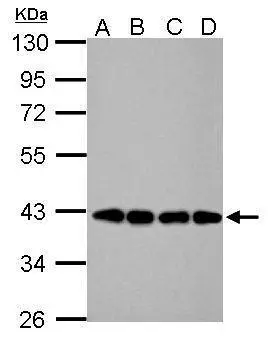
![Various whole cell extracts (30 μg) were separated by 10% SDS-PAGE, and the membrane was blotted with Nanog antibody [GT3312] (GTX627421) diluted at 1:500. The HRP-conjugated anti-mouse IgG antibody (GTX213111-01) was used to detect the primary antibody.](https://www.genetex.com/upload/website/prouct_img/normal/GTX627421/GTX627421_40878_20190607_WB_M_w_23061202_679.webp "Various whole cell extracts (30 μg) were separated by 10% SDS-PAGE, and the membrane was blotted with Nanog antibody [GT3312] (GTX627421) diluted at 1:500. The HRP-conjugated anti-mouse IgG antibody (GTX213111-01) was used to detect the primary antibody.")
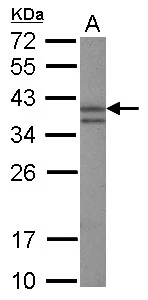
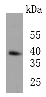
![NANOG antibody [GT3312] detects NANOG protein by western blot analysis. A. 30 μg human ESC whole cell lysate/extract 10% SDS-PAGE NANOG antibody [GT3312] (GTX627421) dilution: 1:1000 The HRP-conjugated anti-mouse IgG antibody (GTX213111-01) was used to detect the primary antibody.](https://www.genetex.com/upload/website/prouct_img/normal/GTX627421/GTX627421_40878_WB_hESC_w_23061202_951.webp "NANOG antibody [GT3312] detects NANOG protein by western blot analysis. A. 30 μg human ESC whole cell lysate/extract 10% SDS-PAGE NANOG antibody [GT3312] (GTX627421) dilution: 1:1000 The HRP-conjugated anti-mouse IgG antibody (GTX213111-01) was used to detect the primary antibody.")
![NANOG antibody [GT3312] detects NANOG protein by flow cytomertry analysis. Sample: Human embryonic stem cells Black: Isotype control dilution: 1:50 Green: NANOG antibody [GT3312] dilution: 1:50](https://www.genetex.com/upload/website/prouct_img/normal/GTX627421/GTX627421_40878_CT_FACS_w_23061202_259.webp "NANOG antibody [GT3312] detects NANOG protein by flow cytomertry analysis. Sample: Human embryonic stem cells Black: Isotype control dilution: 1:50 Green: NANOG antibody [GT3312] dilution: 1:50")
 A: mouse ESC 10% SDS PAGE GTX627421 diluted at 1:1000 The HRP-conjugated anti-mouse IgG antibody (GTX213111-01) was used to detect the primary antibody.")
 in 2019.")

Sample (30 μg of whole cell lysate) A: 293T B: A431 C: HeLa D: HepG2 10% SDS PAGE GTX627421 diluted at 1:1000 The HRP-conjugated anti-mouse IgG antibody (GTX213111-01) was used to detect the primary antibody.
Nanog antibody [GT3312]
GTX627421
ApplicationsFlow Cytometry, ImmunoFluorescence, Western Blot, ImmunoCytoChemistry
Product group Antibodies
ReactivityHuman, Mouse
TargetNANOG
Overview
- SupplierGeneTex
- Product NameNanog antibody [GT3312]
- Delivery Days Customer9
- Application Supplier NoteWB: 1:500-1:3000. FACS: 1:50-1:200. *Optimal dilutions/concentrations should be determined by the researcher.Not tested in other applications.
- ApplicationsFlow Cytometry, ImmunoFluorescence, Western Blot, ImmunoCytoChemistry
- CertificationResearch Use Only
- ClonalityMonoclonal
- Clone IDGT3312
- Concentration1 mg/ml
- ConjugateUnconjugated
- Gene ID79923
- Target nameNANOG
- Target descriptionNanog homeobox
- Target synonymshomeobox protein NANOG, homeobox transcription factor Nanog, homeobox transcription factor Nanog-delta 48
- HostMouse
- IsotypeIgG2b
- Protein IDQ9H9S0
- Protein NameHomeobox protein NANOG
- Scientific DescriptionTranscription regulator involved in inner cell mass and embryonic stem (ES) cells proliferation and self-renewal. Imposes pluripotency on ES cells and prevents their differentiation towards extraembryonic endoderm and trophectoderm lineages. Blocks bone morphogenetic protein-induced mesoderm differentiation of ES cells by physically interacting with SMAD1 and interfering with the recruitment of coactivators to the active SMAD transcriptional complexes (By similarity). Acts as a transcriptional activator or repressor (By similarity). Binds optimally to the DNA consensus sequence 5-TAAT[GT][GT]-3 or 5-[CG][GA][CG]C[GC]ATTAN[GC]-3 (By similarity). When overexpressed, promotes cells to enter into S phase and proliferation.
- ReactivityHuman, Mouse
- Storage Instruction-20°C or -80°C,2°C to 8°C
- UNSPSC12352203
References
- Ye P, Chi X, Yan X, et al. Alanine-Glyoxylate Aminotransferase Sustains Cancer Stemness Properties through the Upregulation of SOX2 and OCT4 in Hepatocellular Carcinoma Cells. Biomolecules. 2022,12(5). doi: 10.3390/biom12050668Read this paper
- Hassan G, Zahra MH, Seno A, et al. The significance of ErbB2/3 in the conversion of induced pluripotent stem cells into cancer stem cells. Sci Rep. 2022,12(1):2711. doi: 10.1038/s41598-022-04980-yRead this paper
- Ciarpella F, Zamfir RG, Campanelli A, et al. Murine cerebral organoids develop network of functional neurons and hippocampal brain region identity. iScience. 2021,24(12):103438. doi: 10.1016/j.isci.2021.103438Read this paper
- Biagioni A, Chillà A, Del Rosso M, et al. CRISPR/Cas9 uPAR Gene Knockout Results in Tumor Growth Inhibition, EGFR Downregulation and Induction of Stemness Markers in Melanoma and Colon Carcinoma Cell Lines. Front Oncol. 2021,11:663225. doi: 10.3389/fonc.2021.663225Read this paper
- Walker SJ, Wagoner AL, Leavitt D, et al. A simplified approach for derivation of induced pluripotent stem cells from Epstein-Barr virus immortalized B-lymphoblastoid cell lines. Heliyon. 2021,7(4):e06617. doi: 10.1016/j.heliyon.2021.e06617Read this paper
- Merle C, Lagarde P, Lartigue L, et al. Acquisition of cancer stem cell capacities after spontaneous cell fusion. BMC Cancer. 2021,21(1):241. doi: 10.1186/s12885-021-07979-2Read this paper
- Chen YL, Yen YC, Jang CW, et al. Ephrin A4-ephrin receptor A10 signaling promotes cell migration and spheroid formation by upregulating NANOG expression in oral squamous cell carcinoma cells. Sci Rep. 2021,11(1):644. doi: 10.1038/s41598-020-80060-3Read this paper
- Lee KY, Kuo TC, Chou CM, et al. Upregulation of CD109 Promotes the Epithelial-to-Mesenchymal Transition and Stemness Properties of Lung Adenocarcinomas via Activation of the Hippo-YAP Signaling. Cells. 2020,10(1). doi: 10.3390/cells10010028Read this paper
- Jao TM, Fang WH, Ciou SC, et al. PCDH10 exerts tumor-suppressor functions through modulation of EGFR/AKT axis in colorectal cancer. Cancer Lett. 2021,499:290-300. doi: 10.1016/j.canlet.2020.11.017Read this paper
- Blanas A, Zaal A, van der Haar Àvila I, et al. FUT9-Driven Programming of Colon Cancer Cells towards a Stem Cell-Like State. Cancers (Basel). 2020,12(9). doi: 10.3390/cancers12092580Read this paper
Datasheet
Related products
Product group Antibodies
Anti-Nanog Antibody118-10023
ApplicationsELISA, ImmunoHistoChemistry
ReactivityHuman
- SizePrice

Product group Antibodies
Anti-Nanog Antibody Picoband(r)A00153-3-CARRIER-FREE
ApplicationsFlow Cytometry, Western Blot
ReactivityHuman, Mouse, Rat
TargetNANOG
- SizePrice
Product group Antibodies
References
Nanog antibody [N3C3]GTX100863
ApplicationsImmunoFluorescence, ImmunoPrecipitation, Western Blot, ELISA, ImmunoCytoChemistry, ImmunoHistoChemistry, ImmunoHistoChemistry Frozen, ImmunoHistoChemistry Paraffin
ReactivityHuman, Mammals, Mouse
TargetNANOG
- SizePrice
Product group Antibodies
Nanog antibody [5A10]GTX57553
ApplicationsFlow Cytometry, Western Blot, ImmunoHistoChemistry, ImmunoHistoChemistry Paraffin
ReactivityHuman, Mouse
TargetNANOG
- SizePrice
Product group Antibodies
Anti-Nanog AntibodyA85213
ApplicationsWestern Blot, ELISA
ReactivityMouse
- SizePrice
![WB analysis of NTERA-2 cell lysate using GTX83107 Nanog antibody [1E6C4].](https://www.genetex.com/upload/website/prouct_img/normal/GTX83107/GTX83107_20170912_WB_w_23061322_951.webp)
Product group Antibodies
Nanog antibody [1E6C4]GTX83107
ApplicationsImmunoFluorescence, Western Blot, ELISA, ImmunoCytoChemistry
ReactivityHuman, Mouse
TargetNANOG
- SizePrice
Product group Antibodies
Nanog Polyclonal AntibodyCAC09911
ApplicationsWestern Blot, ELISA, ImmunoHistoChemistry
TargetNANOG
- SizePrice
Product group Antibodies
Nanog Recombinant AntibodyBSM-52124R
ApplicationsImmunoFluorescence, Western Blot, ImmunoCytoChemistry, ImmunoHistoChemistry, ImmunoHistoChemistry Frozen, ImmunoHistoChemistry Paraffin
ReactivityHuman, Mouse, Rat
TargetNANOG
- SizePrice
Product group Antibodies
Nanog antibody, C-termGTX89079
ApplicationsWestern Blot, ELISA, ImmunoHistoChemistry, ImmunoHistoChemistry Paraffin
ReactivityHuman
TargetNANOG
- SizePrice