

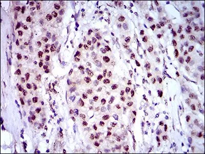
IHC-P analysis of liver cancer tissue using GTX60785 Napsin A antibody [10C4B8].
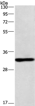
![WB analysis of HEK293 (1) and Napsin A (AA: 20-158)-hIgGFc transfected HEK293 (2) cell lysate using GTX60785 Napsin A antibody [10C4B8].](https://www.genetex.com/upload/website/prouct_img/normal/GTX60785/GTX60785_20170912_WB_w_23061123_235.webp "WB analysis of HEK293 (1) and Napsin A (AA: 20-158)-hIgGFc transfected HEK293 (2) cell lysate using GTX60785 Napsin A antibody [10C4B8].")
![IHC-P analysis of rectum cancer tissue using GTX60785 Napsin A antibody [10C4B8].](https://www.genetex.com/upload/website/prouct_img/normal/GTX60785/GTX60785_20170912_IHC-P_1_w_23061123_345.webp "IHC-P analysis of rectum cancer tissue using GTX60785 Napsin A antibody [10C4B8].")
![ELISA analysis of antigen using GTX60785 Napsin A antibody [10C4B8].
Black : Control antigen 100ng
Purple : Antigen 10ng
Blue : Antigen 50ng
Red : Antigen 100ng](https://www.genetex.com/upload/website/prouct_img/normal/GTX60785/GTX60785_20170912_ELISA_w_23061123_487.webp "ELISA analysis of antigen using GTX60785 Napsin A antibody [10C4B8].
Black : Control antigen 100ng
Purple : Antigen 10ng
Blue : Antigen 50ng
Red : Antigen 100ng")
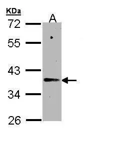
![WB analysis of Rat liver tissue lysate using GTX60785 Napsin A antibody [10C4B8].](https://www.genetex.com/upload/website/prouct_img/normal/GTX60785/GTX60785_20170912_WB_1_w_23061123_179.webp "WB analysis of Rat liver tissue lysate using GTX60785 Napsin A antibody [10C4B8].")
IHC-P analysis of liver cancer tissue using GTX60785 Napsin A antibody [10C4B8].
Napsin A antibody [10C4B8]
GTX60785
ApplicationsWestern Blot, ELISA, ImmunoHistoChemistry, ImmunoHistoChemistry Paraffin
Product group Antibodies
ReactivityHuman, Rat
TargetNAPSA
Overview
- SupplierGeneTex
- Product NameNapsin A antibody [10C4B8]
- Delivery Days Customer9
- Application Supplier NoteWB: 1/500 - 1/2000. IHC-P: 1/200 - 1/1000. ELISA: 1/10000. *Optimal dilutions/concentrations should be determined by the researcher.Not tested in other applications.
- ApplicationsWestern Blot, ELISA, ImmunoHistoChemistry, ImmunoHistoChemistry Paraffin
- CertificationResearch Use Only
- ClonalityMonoclonal
- Clone ID10C4B8
- Concentration1 mg/ml
- ConjugateUnconjugated
- Gene ID9476
- Target nameNAPSA
- Target descriptionnapsin A aspartic peptidase
- Target synonymsKAP, Kdap, NAP1, NAPA, NR1H2-AS1, SNAPA, napsin-A, ASP4, NR1H2 antisense RNA 1, TA01/TA02, asp 4, aspartyl protease 4, kidney-derived aspartic protease-like protein, napsin-1, pronapsin A
- HostMouse
- IsotypeIgG1
- Protein IDO96009
- Protein NameNapsin-A
- Scientific DescriptionThe activation peptides of aspartic proteinases plays role as inhibitors of the active site. These peptide segments, or pro-parts, are deemed important for correct folding, targeting, and control of the activation of aspartic proteinase zymogens. The pronapsin A gene is expressed predominantly in lung and kidney. Its translation product is predicted to be a fully functional, glycosylated aspartic proteinase precursor containing an RGD motif and an additional 18 residues at its C-terminus. [provided by RefSeq, Jul 2008]
- ReactivityHuman, Rat
- Storage Instruction-20°C or -80°C,2°C to 8°C
- UNSPSC12352203
Datasheet
Related products
Product group Antibodies

Anti-NAPSIN A [21A5]Ab03303-10.0
ApplicationsWestern Blot, ELISA, ImmunoHistoChemistry
ReactivityHuman
TargetNAPSA
- SizePrice
Product group Antibodies
Anti-NAPSA Antibody144-65943
ApplicationsWestern Blot
ReactivityHuman, Mouse, Rat
TargetNAPSA
- SizePrice
Product group Antibodies
Napsin A antibody [NAPSA/4400R]GTX02684
ApplicationsImmunoHistoChemistry, ImmunoHistoChemistry Paraffin, Other Application
ReactivityHuman
TargetNAPSA
- SizePrice
![IHC-P analysis of human kidney tissue using GTX04366 Napsin A antibody [MSVA-112R] HistoMAX?. Moderate Napsin A immunostaining in proximal tubuli of the kidney.](https://www.genetex.com/upload/website/prouct_img/normal/GTX04366/GTX04366_20230728_IHC-P_85_23072722_477.webp)
Product group Antibodies
ApplicationsImmunoHistoChemistry, ImmunoHistoChemistry Paraffin
ReactivityHuman
TargetNAPSA
- SizePrice
Product group Antibodies
Napsin A antibody [N1C2]GTX109006
ApplicationsWestern Blot
ReactivityHuman
TargetNAPSA
- SizePrice
Product group Antibodies
NAPSA Polyclonal AntibodyCAC14127
ApplicationsWestern Blot, ELISA, ImmunoHistoChemistry
ReactivityMouse
TargetNAPSA
- SizePrice
Product group Antibodies
Anti-NAPSA AntibodyA37867
ApplicationsWestern Blot, ImmunoHistoChemistry
ReactivityHuman, Mouse
- SizePrice
Product group Antibodies
Napsin A antibody [NAPSA/1238]GTX34887
ApplicationsFlow Cytometry, ImmunoFluorescence, Western Blot, ImmunoCytoChemistry, ImmunoHistoChemistry, ImmunoHistoChemistry Paraffin, Other Application
ReactivityHuman
TargetNAPSA
- SizePrice
Product group Antibodies
NAPSIN A Monoclonal AntibodyBSM-34153M
ApplicationsImmunoFluorescence, ImmunoHistoChemistry, ImmunoHistoChemistry Frozen, ImmunoHistoChemistry Paraffin
ReactivityHuman, Mouse, Rat
TargetNAPSA
- SizePrice