

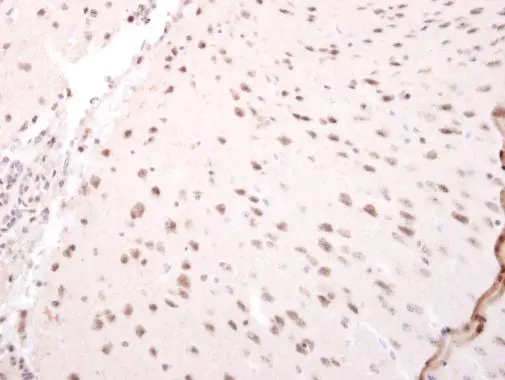
NARF antibody [N3C2], Internal detects NARF protein at nucleus on mouse fore brain by immunohistochemical analysis. Sample: Paraffin-embedded mouse fore brain. NARF antibody [N3C2], Internal (GTX115821) dilution: 1:250.
Antigen Retrieval: Trilogy? (EDTA based, pH 8.0) buffer, 15min
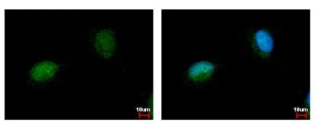
 antibody at 1:500 dilution.")
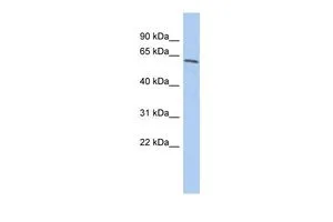
 A: A549 B: MCF-7 7.5% SDS PAGE GTX115821 diluted at 1:1000")
NARF antibody [N3C2], Internal detects NARF protein at nucleus on mouse fore brain by immunohistochemical analysis. Sample: Paraffin-embedded mouse fore brain. NARF antibody [N3C2], Internal (GTX115821) dilution: 1:250.
Antigen Retrieval: Trilogy? (EDTA based, pH 8.0) buffer, 15min
NARF antibody [N3C2], Internal
GTX115821
ApplicationsImmunoFluorescence, Western Blot, ImmunoCytoChemistry, ImmunoHistoChemistry, ImmunoHistoChemistry Paraffin
Product group Antibodies
ReactivityHuman, Mouse
TargetNARF
Overview
- SupplierGeneTex
- Product NameNARF antibody [N3C2], Internal
- Delivery Days Customer9
- Application Supplier NoteWB: 1:500-1:3000. ICC/IF: 1:100-1:1000. IHC-P: 1:100-1:1000. *Optimal dilutions/concentrations should be determined by the researcher.Not tested in other applications.
- ApplicationsImmunoFluorescence, Western Blot, ImmunoCytoChemistry, ImmunoHistoChemistry, ImmunoHistoChemistry Paraffin
- CertificationResearch Use Only
- ClonalityPolyclonal
- Concentration1 mg/ml
- ConjugateUnconjugated
- Gene ID26502
- Target nameNARF
- Target descriptionnuclear prelamin A recognition factor
- Target synonymsIOP2, nuclear prelamin A recognition factor, iron-only hydrogenase-like protein 2, prenyl-dependent prelamin A binding protein
- HostRabbit
- IsotypeIgG
- Protein IDQ9UHQ1
- Protein NameNuclear prelamin A recognition factor
- Scientific DescriptionSeveral proteins have been found to be prenylated and methylated at their carboxyl-terminal ends. Prenylation was initially believed to be important only for membrane attachment. However, another role for prenylation appears to be its importance in protein-protein interactions. The only nuclear proteins known to be prenylated in mammalian cells are prelamin A- and B-type lamins. Prelamin A is farnesylated and carboxymethylated on the cysteine residue of a carboxyl-terminal CaaX motif. This post-translationally modified cysteine residue is removed from prelamin A when it is endoproteolytically processed into mature lamin A. The protein encoded by this gene binds to the prenylated prelamin A carboxyl-terminal tail domain. It may be a component of a prelamin A endoprotease complex. The encoded protein is located in the nucleus, where it partially colocalizes with the nuclear lamina. It shares limited sequence similarity with iron-only bacterial hydrogenases. Alternatively spliced transcript variants encoding different isoforms have been identified for this gene, including one with a novel exon that is generated by RNA editing. [provided by RefSeq]
- ReactivityHuman, Mouse
- Storage Instruction-20°C or -80°C,2°C to 8°C
- UNSPSC41116161
Datasheet
Related products
Product group Antibodies
Anti-NARF (Center) Antibody102-22705
ApplicationsWestern Blot
TargetNARF
- SizePrice
Product group Antibodies
Anti-NARF AntibodyA101363
ApplicationsWestern Blot, ELISA
ReactivityHuman
- SizePrice

Product group Antibodies
Anti-NARF Antibody Picoband(r)A10513-2-CARRIER-FREE
ApplicationsFlow Cytometry, Western Blot, ELISA
ReactivityHuman, Mouse, Rat
TargetNARF
- SizePrice
Product group Antibodies

NARF AntibodyLS-C830513
ApplicationsELISA, ImmunoHistoChemistry
ReactivityHuman, Mouse, Rat
TargetNARF
- SizePrice
Product group Antibodies
NARF Polyclonal AntibodyBS-11173R
ApplicationsImmunoFluorescence, Western Blot, ELISA, ImmunoCytoChemistry, ImmunoHistoChemistry, ImmunoHistoChemistry Frozen, ImmunoHistoChemistry Paraffin
- SizePrice
Product group Antibodies
NARF AntibodyCSB-PA008766
ApplicationsWestern Blot, ELISA
ReactivityHuman
TargetNARF
- SizePrice
Product group Antibodies
NARF Polyclonal AntibodyCAC14858
ApplicationsImmunoFluorescence, Western Blot, ELISA
ReactivityMouse
TargetNARF
- SizePrice

Product group Antibodies
Anti-NARF AntibodyHPA053006
ApplicationsWestern Blot, ImmunoCytoChemistry
ReactivityHuman
TargetNARF
- SizePrice
Product group Antibodies
NARF antibodyGTX115913
ApplicationsImmunoFluorescence, Western Blot, ImmunoCytoChemistry, ImmunoHistoChemistry, ImmunoHistoChemistry Paraffin
ReactivityHuman, Mouse
TargetNARF
- SizePrice
Product group Antibodies
NARF antibody, InternalGTX45406
ApplicationsWestern Blot
ReactivityHuman
TargetNARF
- SizePrice