

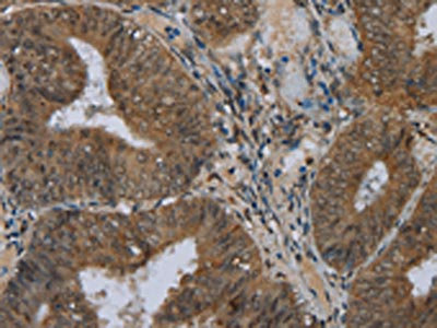
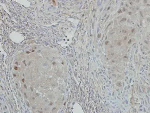
The image on the left is immunohistochemistry of paraffin-embedded Human colon cancer tissue using CSB-PA088333(NAT10 Antibody) at dilution 1/40, on the right is treated with fusion protein. (Original magnification: x200)
 at dilution 1/40, on the right is treated with fusion protein. (Original magnification: x200)")
 at dilution 1/200, Secondary antibody: Goat anti rabbit IgG at 1/8000 dilution, Exposure time: 10 seconds")
The image on the left is immunohistochemistry of paraffin-embedded Human colon cancer tissue using CSB-PA088333(NAT10 Antibody) at dilution 1/40, on the right is treated with fusion protein. (Original magnification: x200)
NAT10 Antibody
CSB-PA088333
ApplicationsWestern Blot, ELISA, ImmunoHistoChemistry
Product group Antibodies
ReactivityHuman, Mouse
TargetNAT10
Overview
- SupplierCusabio
- Product NameNAT10 Antibody
- Delivery Days Customer20
- ApplicationsWestern Blot, ELISA, ImmunoHistoChemistry
- CertificationResearch Use Only
- ClonalityPolyclonal
- ConjugateUnconjugated
- Gene ID55226
- Target nameNAT10
- Target descriptionN-acetyltransferase 10
- Target synonymsALP, Kre33, NET43, RNA cytidine acetyltransferase, 18S rRNA cytosine acetyltransferase, N-acetyltransferase 10 (GCN5-related), N-acetyltransferase-like protein
- HostRabbit
- IsotypeIgG
- Protein IDQ9H0A0
- Protein NameRNA cytidine acetyltransferase
- Scientific DescriptionN-acetyltransferase 10 is an enzyme that in humans is encoded by the NAT10 gene. Has protein acetyltransferase activity in vitro. Can acetylate both histones and microtubules. Histone acetylation may regulate transcription and mitotic chromosome de-condensation. Activates telomerase activity by stimulating the transcription of TERT, and may also regulate telomerase function by affecting the balance of telomerase subunit assembly, disassembly, and localization.
- ReactivityHuman, Mouse
- Storage Instruction-20°C or -80°C
- UNSPSC41116161
Related products
Product group Antibodies
NAT10 Recombinant AntibodyBSM-62424R
ApplicationsFlow Cytometry, ImmunoFluorescence, ImmunoPrecipitation, Western Blot, ImmunoCytoChemistry, ImmunoHistoChemistry, ImmunoHistoChemistry Frozen, ImmunoHistoChemistry Paraffin
ReactivityHuman, Mouse, Rat
TargetNAT10
- SizePrice
Product group Antibodies
Anti-NAT10 Antibody144-66352
ApplicationsImmunoFluorescence, Western Blot
ReactivityHuman
TargetNAT10
- SizePrice

Product group Antibodies
Anti-NAT10 Antibody Picoband(r)A06226-2-CARRIER-FREE
ApplicationsFlow Cytometry, ImmunoFluorescence, ImmunoPrecipitation, Western Blot, ELISA, ImmunoCytoChemistry
ReactivityHuman
TargetNAT10
- SizePrice
Product group Antibodies
Anti-NAT10 AntibodyA31982
ApplicationsImmunoFluorescence, Western Blot, ImmunoHistoChemistry
ReactivityHuman, Mouse, Rat
- SizePrice

Product group Antibodies
Anti-NAT10 AntibodyHPA057576
ApplicationsImmunoCytoChemistry
ReactivityHuman
TargetNAT10
- SizePrice
Product group Antibodies

Goat anti-NAT10 / hALPEB07698
ApplicationsELISA, ImmunoHistoChemistry
ReactivityCanine, Human, Mouse, Rat
TargetNAT10
- SizePrice
Product group Antibodies

NAT10 AntibodyLS-C401867
ApplicationsWestern Blot, ELISA, ImmunoHistoChemistry
ReactivityHuman, Mouse
TargetNAT10
- SizePrice
Product group Antibodies
NAT10 antibody [N1N2], N-termGTX119166
ApplicationsImmunoFluorescence, Western Blot, ImmunoCytoChemistry, ImmunoHistoChemistry, ImmunoHistoChemistry Paraffin
ReactivityHuman
TargetNAT10
- SizePrice