

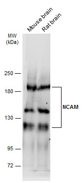
Various tissue extracts (50 μg) were separated by 5% SDS-PAGE, and the membrane was blotted with NCAM antibody (GTX111684) diluted at 1:10000. The HRP-conjugated anti-rabbit IgG antibody (GTX213110-01) was used to detect the primary antibody, and the signal was developed with Trident ECL plus-Enhanced.
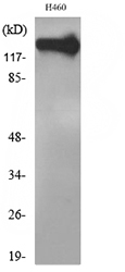
 was separated by 7.5% SDS-PAGE, and the membrane was blotted with NCAM antibody (GTX111684) diluted at 1:10000. The HRP-conjugated anti-rabbit IgG antibody (GTX213110-01) was used to detect the primary antibody.")
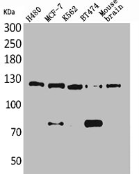
 was separated by 5% SDS-PAGE, and the membrane was blotted with NCAM antibody (GTX111684) diluted at 1:1000. The HRP-conjugated anti-rabbit IgG antibody (GTX213110-01) was used to detect the primary antibody.")
 diluted at 1:100. Blue: Fluoroshield with DAPI (GTX30920).")
 and transfected (+) unboiled 293T whole cell extracts (50 μg) were separated by 7.5% SDS-PAGE, and the membrane was blotted with NCAM antibody (GTX111684) diluted at 1:5000. The HRP-conjugated anti-rabbit IgG antibody (GTX213110-01) was used to detect the primary antibody.")
 were separated by 7.5% SDS-PAGE, and the membrane was blotted with NCAM antibody (GTX111684) diluted at 1:1000. The HRP-conjugated anti-rabbit IgG antibody (GTX213110-01) was used to detect the primary antibody. Corresponding RNA expression data are based on Human Protein Atlas program.")
Various tissue extracts (50 μg) were separated by 5% SDS-PAGE, and the membrane was blotted with NCAM antibody (GTX111684) diluted at 1:10000. The HRP-conjugated anti-rabbit IgG antibody (GTX213110-01) was used to detect the primary antibody, and the signal was developed with Trident ECL plus-Enhanced.
NCAM antibody
GTX111684
ApplicationsImmunoFluorescence, Western Blot, ImmunoCytoChemistry
Product group Antibodies
ReactivityHuman, Mouse, Rat
TargetNCAM1
Overview
- SupplierGeneTex
- Product NameNCAM antibody
- Delivery Days Customer9
- Application Supplier NoteWB: 1:5000-1:20000. *Optimal dilutions/concentrations should be determined by the researcher.Not tested in other applications.
- ApplicationsImmunoFluorescence, Western Blot, ImmunoCytoChemistry
- CertificationResearch Use Only
- ClonalityPolyclonal
- Concentration1 mg/ml
- ConjugateUnconjugated
- Gene ID4684
- Target nameNCAM1
- Target descriptionneural cell adhesion molecule 1
- Target synonymsCD56, MSK39, NCAM, neural cell adhesion molecule 1, antigen recognized by monoclonal antibody 5.1H11, neural cell adhesion molecule, NCAM
- HostRabbit
- IsotypeIgG
- Protein IDP13591
- Protein NameNeural cell adhesion molecule 1
- Scientific DescriptionThis gene encodes a cell adhesion protein which is a member of the immunoglobulin superfamily. The encoded protein is involved in cell-to-cell interactions as well as cell-matrix interactions during development and differentiation. The encoded protein has been shown to be involved in development of the nervous system, and for cells involved in the expansion of T cells and dendritic cells which play an important role in immune surveillance. Alternative splicing results in multiple transcript variants. [provided by RefSeq, Jun 2011]
- ReactivityHuman, Mouse, Rat
- Storage Instruction-20°C or -80°C,2°C to 8°C
- UNSPSC41116161
Datasheet
Related products
Product group Antibodies
NCAM1 AntibodyCSB-PA004909
ApplicationsWestern Blot, ELISA
ReactivityHuman
TargetNCAM1
- SizePrice

Product group Antibodies
Anti-NCAM1 Antibody Picoband(r)A00184-4-CARRIER-FREE
ApplicationsFlow Cytometry, ImmunoFluorescence, Western Blot, ELISA, ImmunoCytoChemistry
ReactivityHuman, Mouse, Rat
TargetNCAM1
- SizePrice
Product group Antibodies
Anti-NCAM1 AntibodyA100955
ApplicationsWestern Blot, ELISA
ReactivityHuman
- SizePrice
Product group Antibodies
ApplicationsFlow Cytometry
ReactivityHuman
TargetNCAM1
- SizePrice
Product group Antibodies
ApplicationsFlow Cytometry
ReactivityHuman
TargetNCAM1
- SizePrice

Product group Antibodies
Anti-NCAM1 AntibodyAMAB91807
ApplicationsImmunoHistoChemistry
ReactivityHuman
TargetNCAM1
- SizePrice
Product group Antibodies

Anti-CD56 [NCAM12.19]Ab01426-1.1
ApplicationsImmunoFluorescence, Western Blot, ELISA
ReactivityHuman
TargetNCAM1
- SizePrice
Product group Antibodies

ApplicationsFlow Cytometry, Western Blot, ELISA, ImmunoHistoChemistry
ReactivityBovine, Human, Porcine, Rat
TargetNCAM1
- SizePrice
Product group Antibodies

NCAM / CD56 AntibodyLS-C406288
ApplicationsELISA, ImmunoHistoChemistry
ReactivityHuman, Mouse, Rat
TargetNCAM1
- SizePrice