

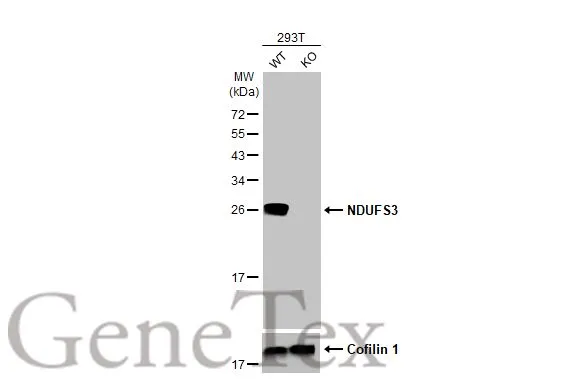
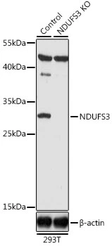
Wild-type (WT) and NDUFS3 knockout (KO) 293T cell extracts (30 μg) were separated by 12% SDS-PAGE, and the membrane was blotted with NDUFS3 antibody [HL2097] (GTX638005) diluted at 1:2500. The HRP-conjugated anti-rabbit IgG antibody (GTX213110-01) was used to detect the primary antibody.
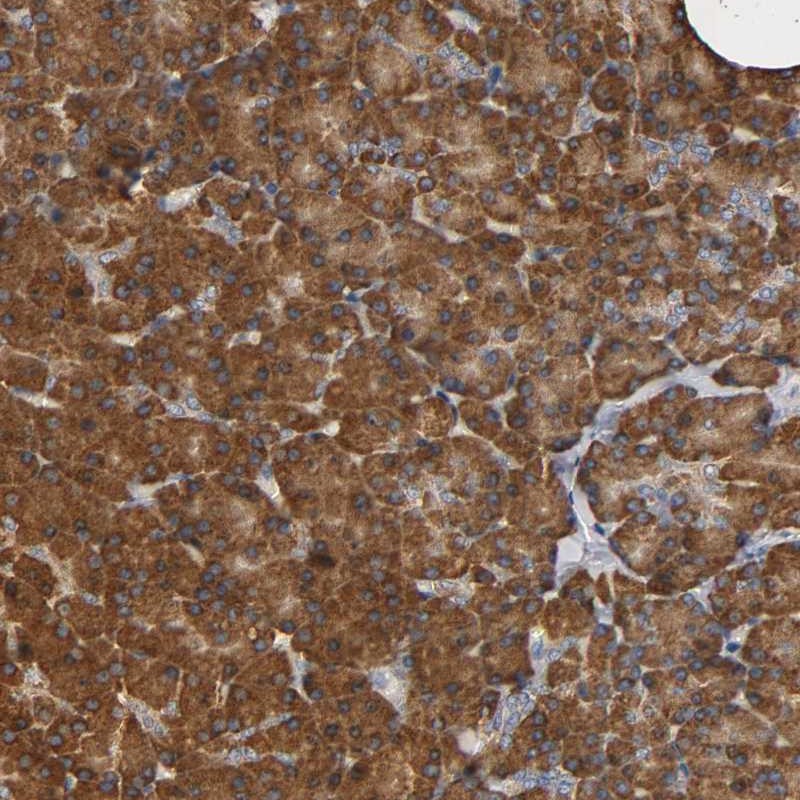
![NDUFS3 antibody [HL2097] detects NDUFS3 protein by immunohistochemical analysis. Sample: Paraffin-embedded mouse tissues. NDUFS3 stained by NDUFS3 antibody [HL2097] (GTX638005) diluted at 1:50. Antigen Retrieval: Citrate buffer, pH 6.0, 15 min](https://www.genetex.com/upload/website/prouct_img/normal/GTX638005/GTX638005_T-44900_20230324_IHC-P_multiple_M_23032819_596.webp "NDUFS3 antibody [HL2097] detects NDUFS3 protein by immunohistochemical analysis. Sample: Paraffin-embedded mouse tissues. NDUFS3 stained by NDUFS3 antibody [HL2097] (GTX638005) diluted at 1:50. Antigen Retrieval: Citrate buffer, pH 6.0, 15 min")
![NDUFS3 antibody [HL2097] detects NDUFS3 protein by immunohistochemical analysis. Sample: Paraffin-embedded rat tissues. NDUFS3 stained by NDUFS3 antibody [HL2097] (GTX638005) diluted at 1:50. Antigen Retrieval: Citrate buffer, pH 6.0, 15 min](https://www.genetex.com/upload/website/prouct_img/normal/GTX638005/GTX638005_T-44900_20230324_IHC-P_multiple_R_23032819_600.webp "NDUFS3 antibody [HL2097] detects NDUFS3 protein by immunohistochemical analysis. Sample: Paraffin-embedded rat tissues. NDUFS3 stained by NDUFS3 antibody [HL2097] (GTX638005) diluted at 1:50. Antigen Retrieval: Citrate buffer, pH 6.0, 15 min")
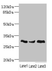
![Various whole cell extracts (30 μg) were separated by 12% SDS-PAGE, and the membrane was blotted with NDUFS3 antibody [HL2097] (GTX638005) diluted at 1:10000. The HRP-conjugated anti-rabbit IgG antibody (GTX213110-01) was used to detect the primary antibody.](https://www.genetex.com/upload/website/prouct_img/normal/GTX638005/GTX638005_44998_20230331_WB_23041023_985.webp "Various whole cell extracts (30 μg) were separated by 12% SDS-PAGE, and the membrane was blotted with NDUFS3 antibody [HL2097] (GTX638005) diluted at 1:10000. The HRP-conjugated anti-rabbit IgG antibody (GTX213110-01) was used to detect the primary antibody.")
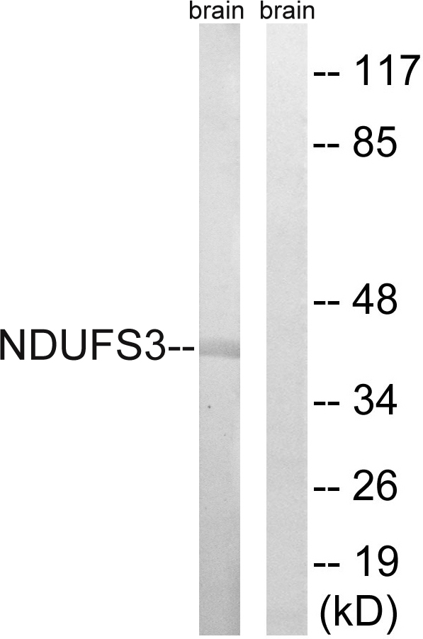
![Various whole cell extracts (30 μg) were separated by 12% SDS-PAGE, and the membrane was blotted with NDUFS3 antibody [HL2097] (GTX638005) diluted at 1:2500. The HRP-conjugated anti-rabbit IgG antibody (GTX213110-01) was used to detect the primary antibody.](https://www.genetex.com/upload/website/prouct_img/normal/GTX638005/GTX638005_T-44900_20230324_WB_23041023_405.webp "Various whole cell extracts (30 μg) were separated by 12% SDS-PAGE, and the membrane was blotted with NDUFS3 antibody [HL2097] (GTX638005) diluted at 1:2500. The HRP-conjugated anti-rabbit IgG antibody (GTX213110-01) was used to detect the primary antibody.")
![NDUFS3 antibody [HL2097] detects NDUFS3 protein at mitochondria by immunofluorescent analysis. Sample: 293T cells were fixed in ice-cold MeOH for 5 min. Green: NDUFS3 stained by NDUFS3 antibody [HL2097] (GTX638005) diluted at 1:500. Red: alpha Tubulin, a cytoskeleton marker, stained by alpha Tubulin antibody [GT114] (GTX628802) diluted at 1:1000. Blue: Fluoroshield with DAPI (GTX30920).](https://www.genetex.com/upload/website/prouct_img/normal/GTX638005/GTX638005_44998_20230602_ICC_IF_23060622_697.webp "NDUFS3 antibody [HL2097] detects NDUFS3 protein at mitochondria by immunofluorescent analysis. Sample: 293T cells were fixed in ice-cold MeOH for 5 min. Green: NDUFS3 stained by NDUFS3 antibody [HL2097] (GTX638005) diluted at 1:500. Red: alpha Tubulin, a cytoskeleton marker, stained by alpha Tubulin antibody [GT114] (GTX628802) diluted at 1:1000. Blue: Fluoroshield with DAPI (GTX30920).")
![Whole zebrafish extract (30 μg) was separated by 12% SDS-PAGE, and the membrane was blotted with NDUFS3 antibody [HL2097] (GTX638005) diluted at 1:1000. The HRP-conjugated anti-rabbit IgG antibody (GTX213110-01) was used to detect the primary antibody.](https://www.genetex.com/upload/website/prouct_img/normal/GTX638005/GTX638005_44998_20230609_WB_Z_23061400_677.webp "Whole zebrafish extract (30 μg) was separated by 12% SDS-PAGE, and the membrane was blotted with NDUFS3 antibody [HL2097] (GTX638005) diluted at 1:1000. The HRP-conjugated anti-rabbit IgG antibody (GTX213110-01) was used to detect the primary antibody.")
![Whole Japanese medaka extract (30 μg) was separated by 12% SDS-PAGE, and the membrane was blotted with NDUFS3 antibody [HL2097] (GTX638005) diluted at 1:1000. The HRP-conjugated anti-rabbit IgG antibody (GTX213110-01) was used to detect the primary antibody.](https://www.genetex.com/upload/website/prouct_img/normal/GTX638005/GTX638005_44998_20250815_WB_medaka_25082121_535.webp "Whole Japanese medaka extract (30 μg) was separated by 12% SDS-PAGE, and the membrane was blotted with NDUFS3 antibody [HL2097] (GTX638005) diluted at 1:1000. The HRP-conjugated anti-rabbit IgG antibody (GTX213110-01) was used to detect the primary antibody.")
Wild-type (WT) and NDUFS3 knockout (KO) 293T cell extracts (30 μg) were separated by 12% SDS-PAGE, and the membrane was blotted with NDUFS3 antibody [HL2097] (GTX638005) diluted at 1:2500. The HRP-conjugated anti-rabbit IgG antibody (GTX213110-01) was used to detect the primary antibody.
NDUFS3 antibody [HL2097]
GTX638005
ApplicationsImmunoFluorescence, Western Blot, ImmunoCytoChemistry, ImmunoHistoChemistry, ImmunoHistoChemistry Paraffin
Product group Antibodies
ReactivityHuman, Mouse, Rat, Zebra Fish
TargetNDUFS3
Overview
- SupplierGeneTex
- Product NameNDUFS3 antibody [HL2097]
- Delivery Days Customer9
- Application Supplier NoteWB: 1:500-1:3000. *Optimal dilutions/concentrations should be determined by the researcher.Not tested in other applications.
- ApplicationsImmunoFluorescence, Western Blot, ImmunoCytoChemistry, ImmunoHistoChemistry, ImmunoHistoChemistry Paraffin
- CertificationResearch Use Only
- ClonalityMonoclonal
- Clone IDHL2097
- Concentration0.5 mg/ml
- ConjugateUnconjugated
- Gene ID4722
- Target nameNDUFS3
- Target descriptionNADH:ubiquinone oxidoreductase core subunit S3
- Target synonymsCI-30, MC1DN8, NADH dehydrogenase [ubiquinone] iron-sulfur protein 3, mitochondrial, CI-30kD, NADH dehydrogenase (ubiquinone) Fe-S protein 3, 30kDa (NADH-coenzyme Q reductase), NADH dehydrogenase-ubiquinone 30 kDa subunit, NADH-ubiquinone oxidoreductase 30 kDa subunit, complex I 30kDa subunit, complex I-30kD
- HostRabbit
- IsotypeIgG
- Protein IDO75489
- Protein NameNADH dehydrogenase [ubiquinone] iron-sulfur protein 3, mitochondrial
- Scientific DescriptionThis gene encodes one of the iron-sulfur protein (IP) components of mitochondrial NADH:ubiquinone oxidoreductase (complex I). Mutations in this gene are associated with Leigh syndrome resulting from mitochondrial complex I deficiency.[provided by RefSeq, Apr 2009]
- ReactivityHuman, Mouse, Rat, Zebra Fish
- Storage Instruction-20°C or -80°C,2°C to 8°C
- UNSPSC41116161
Datasheet
Related products
Product group Antibodies
NDUFS3 AntibodyCSB-PA015662ESR1HU
ApplicationsImmunoFluorescence, Western Blot, ELISA, ImmunoHistoChemistry
ReactivityHuman, Mouse
TargetNDUFS3
- SizePrice
Product group Antibodies
Ndufs3 Recombinant AntibodyCAC12382
ApplicationsWestern Blot, ELISA, ImmunoHistoChemistry
TargetNDUFS3
- SizePrice
Product group Antibodies
Anti-NDUFS3 Antibody144-08013
ApplicationsWestern Blot, ImmunoHistoChemistry
ReactivityHuman, Mouse, Rat
TargetNDUFS3
- SizePrice
Product group Antibodies
Anti-NDUFS3 AntibodyA98781
ApplicationsWestern Blot, ELISA
ReactivityHuman, Mouse
- SizePrice
Product group Antibodies

Goat anti-NDUFS3EB09313
ApplicationsWestern Blot, ELISA
ReactivityBovine, Human
TargetNDUFS3
- SizePrice
Product group Antibodies
Anti-NDUFS3 AntibodyHPA004484
ApplicationsWestern Blot, ImmunoCytoChemistry, ImmunoHistoChemistry
ReactivityHuman
TargetNDUFS3
- SizePrice
Product group Antibodies

NDUFS3 AntibodyLS-C401878
ApplicationsWestern Blot, ELISA, ImmunoHistoChemistry
ReactivityHuman, Mouse
TargetNDUFS3
- SizePrice
Product group Antibodies
NDUFS3 antibodyGTX64675
ApplicationsWestern Blot, ImmunoHistoChemistry, ImmunoHistoChemistry Paraffin
ReactivityHuman, Mouse, Rat
TargetNDUFS3
- SizePrice