

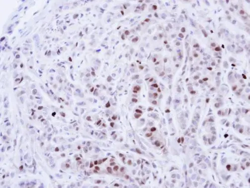
Immunohistochemical analysis of paraffin-embedded A549 Xenograft, using NEK4(GTX102826) antibody at 1:100 dilution.
Antigen Retrieval: Citrate buffer, pH 6.0, 15 min
 A: A431 (GTX27909) B: H1299 7.5% SDS PAGE GTX102826 diluted at 1:1000")
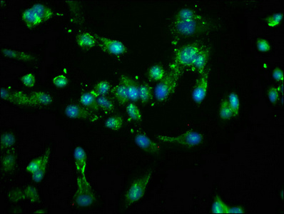
![NEK4 antibody [N2C1], Internal detects NEK4 protein at nucleus by confocal immunofluorescent analysis. Sample: HeLa cells were fixed in ice-cold MeOH for 15 min. Green: NEK4 protein stained by NEK4 antibody [N2C1], Internal (GTX102826) diluted at 1:500. Blue: Hoechst 33343 staining. [Images captured by Olympus FV10i Confocal Laser Scanning Microscope]](https://www.genetex.com/upload/website/prouct_img/normal/GTX102826/GTX102826_40086_IFA_w_23060119_157.webp "NEK4 antibody [N2C1], Internal detects NEK4 protein at nucleus by confocal immunofluorescent analysis. Sample: HeLa cells were fixed in ice-cold MeOH for 15 min. Green: NEK4 protein stained by NEK4 antibody [N2C1], Internal (GTX102826) diluted at 1:500. Blue: Hoechst 33343 staining. [Images captured by Olympus FV10i Confocal Laser Scanning Microscope]")
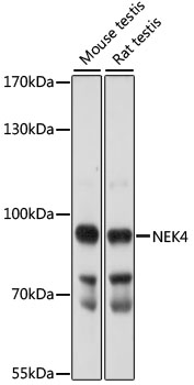
![Various whole cell extracts (30 μg) were separated by 7.5% SDS-PAGE, and the membrane was blotted with NEK4 antibody [N2C1], Internal (GTX102826) diluted at 1:1000. The HRP-conjugated anti-rabbit IgG antibody (GTX213110-01) was used to detect the primary antibody.](https://www.genetex.com/upload/website/prouct_img/normal/GTX102826/GTX102826_40086_20180615_WB_w_23060119_341.webp "Various whole cell extracts (30 μg) were separated by 7.5% SDS-PAGE, and the membrane was blotted with NEK4 antibody [N2C1], Internal (GTX102826) diluted at 1:1000. The HRP-conjugated anti-rabbit IgG antibody (GTX213110-01) was used to detect the primary antibody.")
Immunohistochemical analysis of paraffin-embedded A549 Xenograft, using NEK4(GTX102826) antibody at 1:100 dilution.
Antigen Retrieval: Citrate buffer, pH 6.0, 15 min
NEK4 antibody [N2C1], Internal
GTX102826
ApplicationsImmunoFluorescence, Western Blot, ImmunoCytoChemistry, ImmunoHistoChemistry, ImmunoHistoChemistry Paraffin
Product group Antibodies
ReactivityHuman
TargetNEK4
Overview
- SupplierGeneTex
- Product NameNEK4 antibody [N2C1], Internal
- Delivery Days Customer9
- Application Supplier NoteWB: 1:500-1:3000. ICC/IF: 1:100-1:1000. IHC-P: 1:100-1:1000. *Optimal dilutions/concentrations should be determined by the researcher.Not tested in other applications.
- ApplicationsImmunoFluorescence, Western Blot, ImmunoCytoChemistry, ImmunoHistoChemistry, ImmunoHistoChemistry Paraffin
- CertificationResearch Use Only
- ClonalityPolyclonal
- Concentration0.37 mg/ml
- ConjugateUnconjugated
- Gene ID6787
- Target nameNEK4
- Target descriptionNIMA related kinase 4
- Target synonymsNRK2, STK2, pp12301, serine/threonine-protein kinase Nek4, NIMA (never in mitosis gene a)-related kinase 4, never in mitosis A-related kinase 4, nimA-related protein kinase 4, serine/threonine kinase 2, serine/threonine protein kinase-2, serine/threonine-protein kinase NRK2
- HostRabbit
- IsotypeIgG
- Protein IDP51957
- Protein NameSerine/threonine-protein kinase Nek4
- ReactivityHuman
- Storage Instruction-20°C or -80°C,2°C to 8°C
- UNSPSC12352203
Datasheet
Related products
Product group Antibodies
Anti-NEK4 (C-term) Antibody102-27491
ApplicationsWestern Blot, ImmunoHistoChemistry, ImmunoHistoChemistry Paraffin
TargetNEK4
- SizePrice
Product group Antibodies
Anti-NEK4 AntibodyA91455
ApplicationsWestern Blot
ReactivityMouse, Rat
- SizePrice
Product group Antibodies
NEK4 AntibodyCSB-PA015702LA01HU
ApplicationsImmunoFluorescence, ELISA
ReactivityHuman
TargetNEK4
- SizePrice
Product group Antibodies
Anti-Nek4 AntibodyA09128
ApplicationsImmunoFluorescence, ELISA, ImmunoHistoChemistry
ReactivityHuman, Mouse, Rat
TargetNEK4
- SizePrice
Product group Antibodies

NEK4 AntibodyLS-C671117
ApplicationsImmunoFluorescence, ELISA
ReactivityHuman
TargetNEK4
- SizePrice

Product group Antibodies
Anti-NEK4 AntibodyHPA015750
ApplicationsImmunoHistoChemistry
ReactivityHuman
TargetNEK4
- SizePrice