

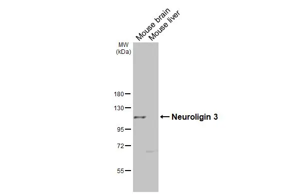
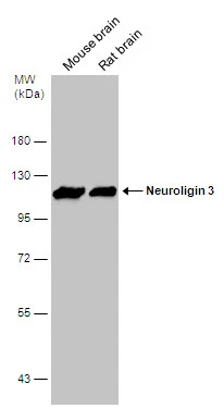
Various tissue extracts (50 μg) were separated by 7.5% SDS-PAGE, and the membrane was blotted with Neuroligin 3 antibody [GT65] (GTX641603) diluted at 1:1000. The HRP-conjugated anti-mouse IgG antibody (GTX213111-01) was used to detect the primary antibody.
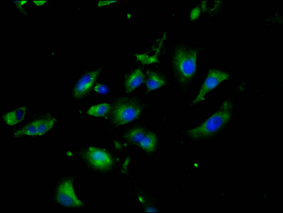
![Neuroligin 3 antibody [GT65] detects Neuroligin 3 protein by immunofluorescent analysis. Sample: DIV9 rat E18 primary hippocampal neuron cells were fixed in 4% paraformaldehyde at RT for 15 min. Green: Neuroligin 3 stained by Neuroligin 3 antibody [GT65] (GTX641603) diluted at 1:250. Red: beta Tubulin 3/ Tuj1 stained by beta Tubulin 3/ Tuj1 antibody (GTX130245) diluted at 1:500. Blue: Fluoroshield with DAPI (GTX30920).](https://www.genetex.com/upload/website/prouct_img/normal/GTX641603/GTX641603_45635_20250509_ICC_IF_R_25052800_712.webp "Neuroligin 3 antibody [GT65] detects Neuroligin 3 protein by immunofluorescent analysis. Sample: DIV9 rat E18 primary hippocampal neuron cells were fixed in 4% paraformaldehyde at RT for 15 min. Green: Neuroligin 3 stained by Neuroligin 3 antibody [GT65] (GTX641603) diluted at 1:250. Red: beta Tubulin 3/ Tuj1 stained by beta Tubulin 3/ Tuj1 antibody (GTX130245) diluted at 1:500. Blue: Fluoroshield with DAPI (GTX30920).")
Various tissue extracts (50 μg) were separated by 7.5% SDS-PAGE, and the membrane was blotted with Neuroligin 3 antibody [GT65] (GTX641603) diluted at 1:1000. The HRP-conjugated anti-mouse IgG antibody (GTX213111-01) was used to detect the primary antibody.
Neuroligin 3 antibody [GT65]
GTX641603
ApplicationsImmunoFluorescence, Western Blot, ImmunoCytoChemistry
Product group Antibodies
ReactivityHuman, Mouse
TargetNLGN3
Overview
- SupplierGeneTex
- Product NameNeuroligin 3 antibody [GT65]
- Delivery Days Customer9
- Application Supplier NoteWB: 1:500-1:3000. *Optimal dilutions/concentrations should be determined by the researcher.Not tested in other applications.
- ApplicationsImmunoFluorescence, Western Blot, ImmunoCytoChemistry
- CertificationResearch Use Only
- ClonalityMonoclonal
- Clone IDGT65
- Concentration1 mg/ml
- ConjugateUnconjugated
- Gene ID54413
- Target nameNLGN3
- Target descriptionneuroligin 3
- Target synonymsHNL3, neuroligin-3, gliotactin homolog
- HostMouse
- IsotypeIgG2b
- Protein IDQ9NZ94
- Protein NameNeuroligin-3
- Scientific DescriptionThis gene encodes a member of a family of neuronal cell surface proteins. Members of this family may act as splice site-specific ligands for beta-neurexins and may be involved in the formation and remodeling of central nervous system synapses. Mutations in this gene may be associated with autism and Asperger syndrome. Multiple transcript variants encoding distinct isoforms have been identified for this gene. [provided by RefSeq, Oct 2009]
- ReactivityHuman, Mouse
- Storage Instruction-20°C or -80°C,2°C to 8°C
- UNSPSC41116161
Related products
Product group Antibodies
Anti-NLGN3 AntibodyA28904
ApplicationsWestern Blot
ReactivityHuman, Mouse
- SizePrice
Product group Antibodies

Goat anti-Neuroligin 3EB08221
ApplicationsWestern Blot, ELISA
ReactivityCanine, Human, Mouse, Rat
TargetNLGN3
- SizePrice

Product group Antibodies
Anti-NLGN3 AntibodyHPA003183
ApplicationsWestern Blot, ImmunoHistoChemistry
ReactivityHuman
TargetNLGN3
- SizePrice
Product group Antibodies
Anti-Neuroligin-3 NLGN3 AntibodyA04627-1
ApplicationsWestern Blot, ELISA
ReactivityHuman, Mouse, Rat
TargetNLGN3
- SizePrice
Product group Antibodies
NLGN3 AntibodyCSB-PA873703LA01HU
ApplicationsImmunoFluorescence, ELISA
ReactivityHuman
TargetNLGN3
- SizePrice
Product group Antibodies

NLGN3 / Neuroligin 3 AntibodyLS-C497018
ApplicationsWestern Blot
ReactivityHuman, Mouse
TargetNLGN3
- SizePrice
![Rat tissue extract (50 μg) was separated by 7.5% SDS-PAGE, and the membrane was blotted with Neuroligin 3 antibody [HL3585] (GTX641556) diluted at 1:1000. The HRP-conjugated anti-rabbit IgG antibody (GTX213110-01) was used to detect the primary antibody.](https://www.genetex.com/upload/website/prouct_img/normal/GTX641556/GTX641556_T-45628_20241220_WB_R_brain_24122400_384.webp)
Product group Antibodies
Neuroligin 3 antibody [HL3585]GTX641556
ApplicationsWestern Blot, ImmunoHistoChemistry, ImmunoHistoChemistry Paraffin
ReactivityHuman, Mouse, Rat
TargetNLGN3
- SizePrice
Product group Antibodies
Neuroligin 3 antibody, InternalGTX88872
ApplicationsWestern Blot
ReactivityHuman
TargetNLGN3
- SizePrice
Product group Antibodies
Neuroligin 3 antibodyGTX131085
ApplicationsImmunoFluorescence, Western Blot, ImmunoCytoChemistry
ReactivityHuman, Mouse, Rat
TargetNLGN3
- SizePrice
Product group Antibodies
Anti-NLGN3 (C-term) Antibody102-24483
ApplicationsWestern Blot
TargetNLGN3
- SizePrice