

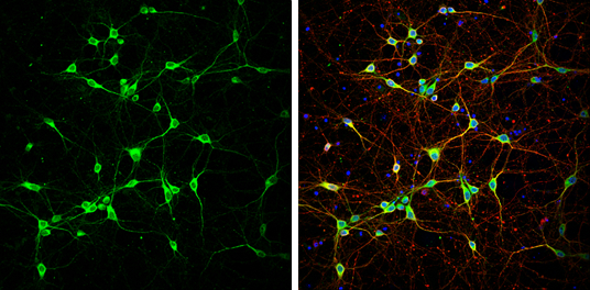
NF-H antibody detects NF-H protein at cytoplasm by immunofluorescent analysis. Sample: DIV9 rat E18 primary cortical neurons were fixed in 4% paraformaldehyde at RT for 15 min. Green: NF-H protein stained by NF-H antibody (GTX110065) diluted at 1:500. Red: beta Tubulin 3/ Tuj1, stained by beta Tubulin 3/ Tuj1 antibody [GT11710] (GTX631836) diluted at 1:500. Blue: Fluoroshield with DAPI (GTX30920).
![NF-H antibody detects NF-H protein expression by immunohistochemical analysis. Sample: Frozen-sectioned adult mouse cerebellum. Green: NF-H protein stained by NF-H antibody (GTX110065) diluted at 1:250. Red: beta Tubulin 3/ TUJ1, stained by beta Tubulin 3/ TUJ1 antibody [GT11710] (GTX631836) diluted at 1:500. Blue: Fluoroshield with DAPI (GTX30920).](https://www.genetex.com/upload/website/prouct_img/normal/GTX110065/GTX110065_40114_20170531_IHC-Fr_M_2_w_23060500_210.webp "NF-H antibody detects NF-H protein expression by immunohistochemical analysis. Sample: Frozen-sectioned adult mouse cerebellum. Green: NF-H protein stained by NF-H antibody (GTX110065) diluted at 1:250. Red: beta Tubulin 3/ TUJ1, stained by beta Tubulin 3/ TUJ1 antibody [GT11710] (GTX631836) diluted at 1:500. Blue: Fluoroshield with DAPI (GTX30920).")
![NF-H antibody detects NF-H protein expression by immunohistochemical analysis. Sample:Paraffin-Embedded adult mouse retina. Green: NF-H protein stained by NF-H antibody (GTX110065) diluted at 1:250. Red: beta Tubulin 3/ TUJ1, stained by beta Tubulin 3/ TUJ1 antibody [GT11710] (GTX631836) diluted at 1:500. Blue: Fluoroshield with DAPI (GTX30920).](https://www.genetex.com/upload/website/prouct_img/normal/GTX110065/GTX110065_40114_20170522_IHC-P_M_w_23060500_498.webp "NF-H antibody detects NF-H protein expression by immunohistochemical analysis. Sample:Paraffin-Embedded adult mouse retina. Green: NF-H protein stained by NF-H antibody (GTX110065) diluted at 1:250. Red: beta Tubulin 3/ TUJ1, stained by beta Tubulin 3/ TUJ1 antibody [GT11710] (GTX631836) diluted at 1:500. Blue: Fluoroshield with DAPI (GTX30920).")
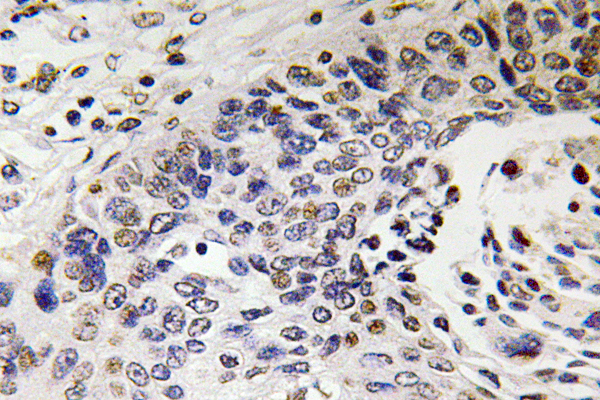
 diluted at 1:500. Antigen Retrieval: Citrate buffer, pH 6.0, 15 min")
 was separated by 5% SDS-PAGE, and the membrane was blotted with NF-H antibody (GTX110065) diluted at 1:500. The HRP-conjugated anti-rabbit IgG antibody (GTX213110-01) was used to detect the primary antibody.")
![NF-H antibody detects NF-H protein expression by immunohistochemical analysis. Sample: Frozen-sectioned adult mouse cerebellum. Green: NF-H protein stained by NF-H antibody (GTX110065) diluted at 1:250. Red: beta Tubulin 3/ TUJ1, stained by beta Tubulin 3/ TUJ1 antibody [GT11710] (GTX631836) diluted at 1:500. Blue: Fluoroshield with DAPI (GTX30920).](https://www.genetex.com/upload/website/prouct_img/normal/GTX110065/GTX110065_40114_20170531_IHC-Fr_M_1_w_23060500_241.webp "NF-H antibody detects NF-H protein expression by immunohistochemical analysis. Sample: Frozen-sectioned adult mouse cerebellum. Green: NF-H protein stained by NF-H antibody (GTX110065) diluted at 1:250. Red: beta Tubulin 3/ TUJ1, stained by beta Tubulin 3/ TUJ1 antibody [GT11710] (GTX631836) diluted at 1:500. Blue: Fluoroshield with DAPI (GTX30920).")
![NF-H antibody detects NF-H protein expression by immunohistochemical analysis. Sample: Frozen sectioned E13.5 Rat brain. Green: NF-H protein stained by NF-H antibody (GTX110065) diluted at 1:250. Red: beta Tubulin 3/ TUJ1, a mature neuron marker, stained by beta Tubulin 3/ TUJ1 antibody [GT11710] (GTX631836) diluted at 1:500. Blue: Fluoroshield with DAPI (GTX30920).](https://www.genetex.com/upload/website/prouct_img/normal/GTX110065/GTX110065_40114_20160921_IHC-Fr_w_23060500_128.webp "NF-H antibody detects NF-H protein expression by immunohistochemical analysis. Sample: Frozen sectioned E13.5 Rat brain. Green: NF-H protein stained by NF-H antibody (GTX110065) diluted at 1:250. Red: beta Tubulin 3/ TUJ1, a mature neuron marker, stained by beta Tubulin 3/ TUJ1 antibody [GT11710] (GTX631836) diluted at 1:500. Blue: Fluoroshield with DAPI (GTX30920).")
 was separated by 5% SDS-PAGE, and the membrane was blotted with NF-H antibody (GTX110065) diluted at 1:500.")
NF-H antibody detects NF-H protein at cytoplasm by immunofluorescent analysis. Sample: DIV9 rat E18 primary cortical neurons were fixed in 4% paraformaldehyde at RT for 15 min. Green: NF-H protein stained by NF-H antibody (GTX110065) diluted at 1:500. Red: beta Tubulin 3/ Tuj1, stained by beta Tubulin 3/ Tuj1 antibody [GT11710] (GTX631836) diluted at 1:500. Blue: Fluoroshield with DAPI (GTX30920).
NF-H antibody
GTX110065
ApplicationsImmunoFluorescence, Western Blot, ImmunoCytoChemistry, ImmunoHistoChemistry, ImmunoHistoChemistry Frozen, ImmunoHistoChemistry Paraffin
Product group Antibodies
ReactivityHuman, Mouse, Rat
TargetNEFH
Overview
- SupplierGeneTex
- Product NameNF-H antibody
- Delivery Days Customer9
- Application Supplier NoteWB: 1:500-1:3000. ICC/IF: 1:100-1:1000. IHC-P: 1:100-1:1000. IHC-Fr: 1:100-1:1000. *Optimal dilutions/concentrations should be determined by the researcher.Not tested in other applications.
- ApplicationsImmunoFluorescence, Western Blot, ImmunoCytoChemistry, ImmunoHistoChemistry, ImmunoHistoChemistry Frozen, ImmunoHistoChemistry Paraffin
- CertificationResearch Use Only
- ClonalityPolyclonal
- Concentration0.71 mg/ml
- ConjugateUnconjugated
- Gene ID4744
- Target nameNEFH
- Target descriptionneurofilament heavy chain
- Target synonymsCMT2CC, NFH, neurofilament heavy polypeptide, 200 kDa neurofilament protein, NF-H, neurofilament triplet H protein, neurofilament, heavy polypeptide 200kDa
- HostRabbit
- IsotypeIgG
- Protein IDP12036
- Protein NameNeurofilament heavy polypeptide
- Scientific DescriptionNeurofilaments are type IV intermediate filament heteropolymers composed of light, medium, and heavy chains. Neurofilaments comprise the axoskeleton and functionally maintain neuronal caliber. They may also play a role in intracellular transport to axons and dendrites. This gene encodes the heavy neurofilament protein. This protein is commonly used as a biomarker of neuronal damage and susceptibility to amyotrophic lateral sclerosis (ALS) has been associated with mutations in this gene. [provided by RefSeq]
- ReactivityHuman, Mouse, Rat
- Storage Instruction-20°C or -80°C,2°C to 8°C
- UNSPSC41116161
Datasheet
Related products
Product group Antibodies
Anti-NF-H AntibodyA101261
ApplicationsELISA, ImmunoHistoChemistry
ReactivityHuman
- SizePrice
Product group Antibodies
Anti-NEFH Antibody144-08442
ApplicationsWestern Blot, ImmunoHistoChemistry
ReactivityHuman, Mouse, Rat
TargetNEFH
- SizePrice

Product group Antibodies
Anti-NEFH AntibodyAMAB91025
ApplicationsWestern Blot, ImmunoHistoChemistry
ReactivityHuman
TargetNEFH
- SizePrice
Product group Antibodies
References
NF-H Polyclonal AntibodyBS-0708R
ApplicationsImmunoFluorescence, Western Blot, ELISA, ImmunoHistoChemistry, ImmunoHistoChemistry Paraffin
ReactivityHuman, Mouse, Rat
TargetNEFH
- SizePrice
Product group Antibodies
ApplicationsImmunoPrecipitation, Western Blot, ImmunoCytoChemistry, ImmunoHistoChemistry
TargetNEFH
- SizePrice
Product group Antibodies
NEFH AntibodyCSB-PA015686LA01HU
ApplicationsImmunoFluorescence, ELISA, ImmunoHistoChemistry
ReactivityHuman
TargetNEFH
- SizePrice
Product group Antibodies

Mouse anti Neurofilament 200 kDMUB1306P
ApplicationsWestern Blot, ImmunoHistoChemistry, ImmunoHistoChemistry Frozen, ImmunoHistoChemistry Paraffin
ReactivityBovine, Canine, Chicken, Guinea Pig, Human, Monkey, Mouse, Rabbit, Rat, Sheep, Xenopus
TargetNEFH
- SizePrice

Product group Antibodies
References
ApplicationsWestern Blot, ImmunoHistoChemistry
ReactivityHuman, Mouse, Rat
TargetNEFH
- SizePrice
Product group Antibodies

NEFH / NF-H AntibodyLS-C406605
ApplicationsELISA, ImmunoHistoChemistry
ReactivityHuman
TargetNEFH
- SizePrice