

NKD1 antibody
70R-3080
Product group Antibodies
Overview
- SupplierBiosynth
- Product NameNKD1 antibody
- Delivery Days Customer7
- CertificationResearch Use Only
- UNSPSC12352203
Related products
Product group Antibodies
NKD1 Polyclonal AntibodyCAC13453
ApplicationsImmunoFluorescence, Western Blot, ELISA
TargetNKD1
- SizePrice
Product group Antibodies
NKD1 AntibodyCSB-PA853369LA01HU
ApplicationsImmunoFluorescence, Western Blot, ELISA
TargetNKD1
- SizePrice

Product group Antibodies
Anti-NKD1 AntibodyHPA049413
ApplicationsImmunoCytoChemistry, ImmunoHistoChemistry
TargetNKD1
- SizePrice
Product group Antibodies
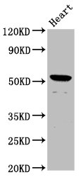
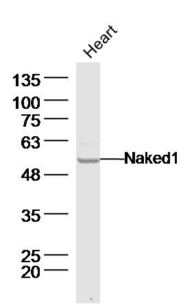
Naked1 Polyclonal AntibodyBS-19005R
ApplicationsImmunoFluorescence, Western Blot, ELISA, ImmunoCytoChemistry, ImmunoHistoChemistry, ImmunoHistoChemistry Frozen, ImmunoHistoChemistry Paraffin
TargetNKD1
- SizePrice

Product group Antibodies
Anti-NKD1 Antibody Picoband(r)A06747-1-CARRIER-FREE
ApplicationsFlow Cytometry, Western Blot, ELISA
TargetNKD1
- SizePrice
Product group Antibodies

NKD1 AntibodyLS-C674467
ApplicationsImmunoFluorescence, Western Blot, ELISA
TargetNKD1
- SizePrice