

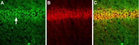
IHC-Fr analysis of mouse brain tissue using GTX03399 NMDAR1 antibody. Staining reveals expression in neurons of the pyramidal layer (an arrow points at the layer). Green : Primary antibody Red : CALHM1 antibody Dilution : 1:200

IHC-Fr analysis of mouse brain tissue using GTX03399 NMDAR1 antibody. Staining reveals expression in neurons of the pyramidal layer (an arrow points at the layer). Green : Primary antibody Red : CALHM1 antibody Dilution : 1:200
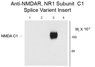
NMDAR1 antibody
GTX03399
ApplicationsImmunoFluorescence, Western Blot, ImmunoCytoChemistry, ImmunoHistoChemistry, ImmunoHistoChemistry Frozen, Other Application
Product group Antibodies
ReactivityHuman, Mouse, Rat
TargetGrin1
Overview
- SupplierGeneTex
- Product NameNMDAR1 antibody
- Delivery Days Customer9
- ApplicationsImmunoFluorescence, Western Blot, ImmunoCytoChemistry, ImmunoHistoChemistry, ImmunoHistoChemistry Frozen, Other Application
- CertificationResearch Use Only
- ClonalityPolyclonal
- Concentration0.8 mg/ml
- ConjugateUnconjugated
- Gene ID24408
- Target nameGrin1
- Target descriptionglutamate ionotropic receptor NMDA type subunit 1
- Target synonymsGluN1, NMDAR1, NR1, glutamate receptor ionotropic, NMDA 1, N-methyl-D-aspartate glutamate receptor, N-methyl-D-aspartate receptor subunit NR1, NMD-R1, NMDA R1 receptor C1 cassette, glutamate [NMDA] receptor subunit zeta-1, glutamate receptor, ionotropic, N-methyl D-aspartate 1, neurotransmitter receptor
- HostRabbit
- IsotypeIgG
- Protein IDP35439
- Protein NameGlutamate receptor ionotropic, NMDA 1
- Scientific Descriptionsubunit of NMDA-preferring ionotropic glutamate receptors; may play a role in long term potentiation [RGD, Feb 2006]
- ReactivityHuman, Mouse, Rat
- Storage Instruction-20°C or -80°C,2°C to 8°C
- UNSPSC12352203
Datasheet
Related products
Product group Antibodies
NMDA NR1 Subunit AntibodyBSM-70179M
ApplicationsWestern Blot, ImmunoHistoChemistry, ImmunoHistoChemistry Paraffin
ReactivityHuman, Mouse, Rat
TargetGrin1
- SizePrice
Product group Antibodies
ApplicationsImmunoPrecipitation, Western Blot, ImmunoCytoChemistry, ImmunoHistoChemistry
ReactivityRat
TargetGrin1
- SizePrice
Product group Antibodies
ApplicationsWestern Blot
ReactivityHuman, Mouse, Rat
TargetGrin1
- SizePrice
Product group Antibodies
ApplicationsWestern Blot, ImmunoHistoChemistry
ReactivityHuman, Mouse, Rat
TargetGrin1
- SizePrice
Product group Antibodies
ApplicationsWestern Blot, ImmunoHistoChemistry
ReactivityHuman, Mouse, Rat
TargetGrin1
- SizePrice
Product group Antibodies
ApplicationsWestern Blot, ImmunoHistoChemistry
ReactivityMouse, Rat
TargetGrin1
- SizePrice