

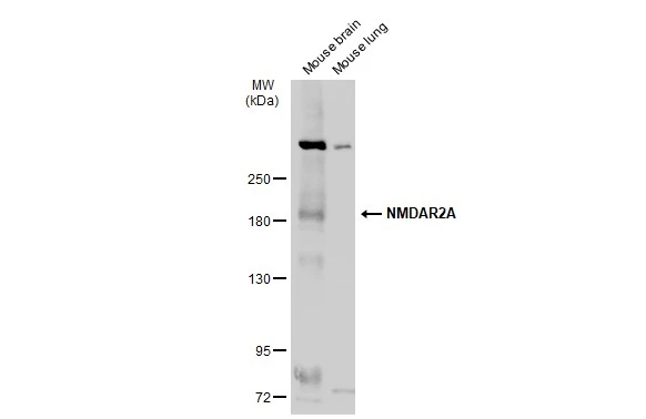
Various tissue extracts (50 μg) were separated by 5% SDS-PAGE, and the membrane was blotted with NMDAR2A antibody (GTX103558) diluted at 1:500. The HRP-conjugated anti-rabbit IgG antibody (GTX213110-01) was used to detect the primary antibody.
 was separated by 5% SDS-PAGE, and the membrane was blotted with NMDAR2A antibody (GTX103558) diluted at 1:500. The HRP-conjugated anti-rabbit IgG antibody (GTX213110-01) was used to detect the primary antibody.")
![NMDAR2A antibody detects NMDAR2A protein at cell membrane by immunofluorescent analysis. Sample: DIV9 rat E18 primary cortical neuron cells were fixed in 4% paraformaldehyde at RT for 15 min. Green: NMDAR2A stained by NMDAR2A antibody (GTX103558) diluted at 1:250. Red: Tau, an axon marker, stained by Tau antibody [GT287] (GTX634809) diluted at 1:500. Blue: Fluoroshield with DAPI (GTX30920).](https://www.genetex.com/upload/website/prouct_img/normal/GTX103558/GTX103558_43915_20221209_ICC_IF_R_22122018_297.webp "NMDAR2A antibody detects NMDAR2A protein at cell membrane by immunofluorescent analysis. Sample: DIV9 rat E18 primary cortical neuron cells were fixed in 4% paraformaldehyde at RT for 15 min. Green: NMDAR2A stained by NMDAR2A antibody (GTX103558) diluted at 1:250. Red: Tau, an axon marker, stained by Tau antibody [GT287] (GTX634809) diluted at 1:500. Blue: Fluoroshield with DAPI (GTX30920).")
 A: Jurkat 5% SDS PAGE GTX103558 diluted at 1:1000 The HRP-conjugated anti-rabbit IgG antibody (GTX213110-01) was used to detect the primary antibody.")
![NMDAR2A antibody detects NMDAR2A protein by immunofluorescent analysis. Sample: DIV14 rat E18 primary cortical neurons were fixed in 4% paraformaldehyde at RT for 15 min. Green: NMDAR2A protein stained by NMDAR2A antibody (GTX103558) diluted at 1:500. Red: beta Tubulin 3/ Tuj1, stained by beta Tubulin 3/ Tuj1 antibody [GT1338] (GTX631831) diluted at 1:500. Blue: Fluoroshield with DAPI (GTX30920).](https://www.genetex.com/upload/website/prouct_img/normal/GTX103558/GTX103558_40394_20170719_IFA_R_w_23060119_258.webp "NMDAR2A antibody detects NMDAR2A protein by immunofluorescent analysis. Sample: DIV14 rat E18 primary cortical neurons were fixed in 4% paraformaldehyde at RT for 15 min. Green: NMDAR2A protein stained by NMDAR2A antibody (GTX103558) diluted at 1:500. Red: beta Tubulin 3/ Tuj1, stained by beta Tubulin 3/ Tuj1 antibody [GT1338] (GTX631831) diluted at 1:500. Blue: Fluoroshield with DAPI (GTX30920).")
 dilution: 1:500.
Antigen Retrieval: Trilogy? (EDTA based, pH 8.0) buffer, 15min")
Various tissue extracts (50 μg) were separated by 5% SDS-PAGE, and the membrane was blotted with NMDAR2A antibody (GTX103558) diluted at 1:500. The HRP-conjugated anti-rabbit IgG antibody (GTX213110-01) was used to detect the primary antibody.
NMDAR2A antibody
GTX103558
ApplicationsImmunoFluorescence, Western Blot, ImmunoCytoChemistry, ImmunoHistoChemistry, ImmunoHistoChemistry Paraffin
Product group Antibodies
ReactivityHuman, Mouse, Rat
TargetGRIN2A
Overview
- SupplierGeneTex
- Product NameNMDAR2A antibody
- Delivery Days Customer9
- Application Supplier NoteWB: 1:500-1:3000. ICC/IF: 1:100-1:1000. IHC-P: 1:100-1:1000. *Optimal dilutions/concentrations should be determined by the researcher.Not tested in other applications.
- ApplicationsImmunoFluorescence, Western Blot, ImmunoCytoChemistry, ImmunoHistoChemistry, ImmunoHistoChemistry Paraffin
- CertificationResearch Use Only
- ClonalityPolyclonal
- Concentration1 mg/ml
- ConjugateUnconjugated
- Gene ID2903
- Target nameGRIN2A
- Target descriptionglutamate ionotropic receptor NMDA type subunit 2A
- Target synonymsEPND, FESD, GluN2A, LKS, NMDAR2A, NR2A, glutamate receptor ionotropic, NMDA 2A, N-methyl D-aspartate receptor subtype 2A, N-methyl-D-aspartate receptor channel, subunit epsilon-1, N-methyl-D-aspartate receptor subunit 2A, glutamate ionotropic receptor NMDA 2A, glutamate receptor, ionotropic, N-methyl D-aspartate 2A
- HostRabbit
- IsotypeIgG
- Protein IDQ12879
- Protein NameGlutamate receptor ionotropic, NMDA 2A
- Scientific DescriptionN-methyl-D-aspartate (NMDA) receptors are a class of ionotropic glutamate-gated ion channels. These receptors have been shown to be involved in long-term potentiation, an activity-dependent increase in the efficiency of synaptic transmission thought to underlie certain kinds of memory and learning. NMDA receptor channels are heteromers composed of the key receptor subunit NMDAR1 (GRIN1) and 1 or more of the 4 NMDAR2 subunits: NMDAR2A (GRIN2A), NMDAR2B (GRIN2B), NMDAR2C (GRIN2C) and NMDAR2D (GRIN2D). Alternatively spliced transcript variants encoding different isoforms have been found for this gene. [provided by RefSeq]
- ReactivityHuman, Mouse, Rat
- Storage Instruction-20°C or -80°C,2°C to 8°C
- UNSPSC41116161
Datasheet
Related products
Product group Antibodies
Anti-NMDAR2A AntibodyA121167
ApplicationsImmunoFluorescence, ELISA
ReactivityHuman, Mouse
- SizePrice
Product group Antibodies
Anti-Mouse/Rat GRIN2A Antibody144-00924
ApplicationsWestern Blot, ImmunoHistoChemistry
ReactivityHuman, Mouse, Rat
TargetGRIN2A
- SizePrice
Product group Antibodies

GRIN2A / NMDAR2A / NR2A AntibodyLS-C748884
ApplicationsImmunoFluorescence, Western Blot
ReactivityHuman, Mouse, Rat
TargetGRIN2A
- SizePrice
Product group Antibodies
References
NMDAR2A Polyclonal AntibodyBS-3507R
ApplicationsWestern Blot, ELISA
ReactivityHuman, Mouse, Rat
TargetGRIN2A
- SizePrice
Product group Antibodies
Phospho-GRIN2A/GRIN2B (Y1246/1252) AntibodyCSB-PA008976
ApplicationsImmunoFluorescence, ELISA, ImmunoHistoChemistry
ReactivityHuman, Mouse, Rat
TargetGRIN2A
- SizePrice
Product group Antibodies

Goat anti-GRIN2A (aa211-224)EB11313
ApplicationsImmunoFluorescence, ELISA
ReactivityCanine, Human, Mouse, Rat
TargetGRIN2A
- SizePrice
Product group Antibodies
Grin2A Polyclonal AntibodyCAC07549
ApplicationsImmunoFluorescence, Western Blot, ELISA, ImmunoHistoChemistry
ReactivityMouse, Rat
TargetGRIN2A
- SizePrice
![NMDAR2A antibody [C1C2], Internal detects GRIN2A protein at cytosol and nucleus on U87 xenograft by immunohistochemical analysis. Sample: Paraffin-embedded U87 xenograft. NMDAR2A antibody [C1C2], Internal (GTX113717) dilution: 1:500.
Antigen Retrieval: Trilogy? (EDTA based, pH 8.0) buffer, 15min](https://www.genetex.com/upload/website/prouct_img/normal/GTX113717/GTX113717_40163_IHC_w_23060501_819.webp)
Product group Antibodies
NMDAR2A antibody [C1C2], InternalGTX113717
ApplicationsImmunoFluorescence, ImmunoCytoChemistry, ImmunoHistoChemistry, ImmunoHistoChemistry Paraffin
ReactivityHuman, Rat
TargetGRIN2A
- SizePrice