

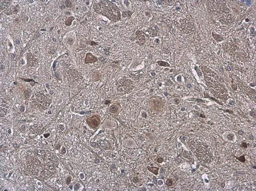
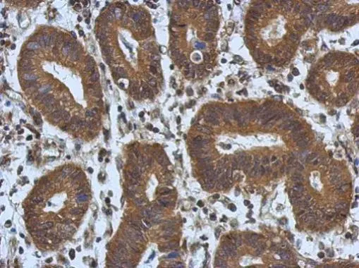
NOL3 antibody detects NOL3 protein at cytoplasm and nucleus by immunohistochemical analysis. Sample: Paraffin-embedded rat brain. NOL3 stained by NOL3 antibody (GTX113889) diluted at 1:500.
Antigen Retrieval: Citrate buffer, pH 6.0, 15 min
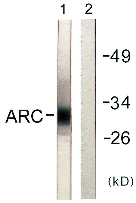
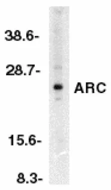
 were separated by 12% SDS-PAGE, and the membrane was blotted with NOL3 antibody (GTX113889) diluted at 1:1000. The HRP-conjugated anti-rabbit IgG antibody (GTX213110-01) was used to detect the primary antibody.")
![NOL3 antibody detects NOL3 protein by immunohistochemical analysis. Sample: Frozen-sectioned mouse cerebellum. Green: NOL3 stained by NOL3 antibody (GTX113889) diluted at 1:250. Red: NF-H, stained by NF-H antibody [GT114] (GTX634289) diluted at 1:500. Blue: Fluoroshield with DAPI (GTX30920).](https://www.genetex.com/upload/website/prouct_img/normal/GTX113889/GTX113889_40660_20180125_IHC-Fr_M_w_23060501_553.webp "NOL3 antibody detects NOL3 protein by immunohistochemical analysis. Sample: Frozen-sectioned mouse cerebellum. Green: NOL3 stained by NOL3 antibody (GTX113889) diluted at 1:250. Red: NF-H, stained by NF-H antibody [GT114] (GTX634289) diluted at 1:500. Blue: Fluoroshield with DAPI (GTX30920).")
 antibody at 1:500 dilution.
Antigen Retrieval: Trilogy? (EDTA based, pH 8.0) buffer, 15min")
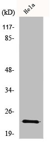
 diluted at 1:500.
Antigen Retrieval: Citrate buffer, pH 6.0, 15 min")
 of paraformaldehyde-fixed HeLa, using NOL3(GTX113889) antibody (Green) at 1:500 dilution. Alpha-tubulin filaments were labeled with GTX11304 (Red) at 1:2000.")
 diluted at 1:500. Antigen Retrieval: Citrate buffer, pH 6.0, 15 min")
 diluted at 1:500. Antigen Retrieval: Citrate buffer, pH 6.0, 15 min")
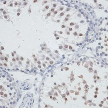
NOL3 antibody detects NOL3 protein at cytoplasm and nucleus by immunohistochemical analysis. Sample: Paraffin-embedded rat brain. NOL3 stained by NOL3 antibody (GTX113889) diluted at 1:500.
Antigen Retrieval: Citrate buffer, pH 6.0, 15 min
NOL3 antibody
GTX113889
ApplicationsImmunoFluorescence, Western Blot, ImmunoCytoChemistry, ImmunoHistoChemistry, ImmunoHistoChemistry Frozen, ImmunoHistoChemistry Paraffin
Product group Antibodies
ReactivityHuman, Mouse, Rat
TargetNOL3
Overview
- SupplierGeneTex
- Product NameNOL3 antibody
- Delivery Days Customer9
- Application Supplier NoteWB: 1:500-1:3000. ICC/IF: 1:100-1:1000. IHC-P: 1:100-1:1000. IHC-Fr: 1:100-1:1000. *Optimal dilutions/concentrations should be determined by the researcher.Not tested in other applications.
- ApplicationsImmunoFluorescence, Western Blot, ImmunoCytoChemistry, ImmunoHistoChemistry, ImmunoHistoChemistry Frozen, ImmunoHistoChemistry Paraffin
- CertificationResearch Use Only
- ClonalityPolyclonal
- Concentration1.3 mg/ml
- ConjugateUnconjugated
- Gene ID8996
- Target nameNOL3
- Target descriptionnucleolar protein 3
- Target synonymsARC, FCM, MYOCL1, MYP, NOP, NOP30, nucleolar protein 3, muscle-enriched cytoplasmic protein, nucleolar protein 3 (apoptosis repressor with CARD domain), nucleolar protein of 30 kDa
- HostRabbit
- IsotypeIgG
- Protein IDO60936
- Protein NameNucleolar protein 3
- Scientific DescriptionThis gene encodes an anti-apoptotic protein that has been shown to down-regulate the enzyme activities of caspase 2, caspase 8 and tumor protein p53. Multiple transcript variants encoding different isoforms have been found for this gene. [provided by RefSeq]
- ReactivityHuman, Mouse, Rat
- Storage Instruction-20°C or -80°C,2°C to 8°C
- UNSPSC41116161
Datasheet
Related products
Product group Antibodies
Anti-ARC AntibodyA100060
ApplicationsWestern Blot, ELISA, ImmunoHistoChemistry
ReactivityHuman
- SizePrice
Product group Antibodies
Anti-NOL3 Antibody144-06319
ApplicationsImmunoFluorescence, Western Blot, ImmunoHistoChemistry
ReactivityHuman, Mouse, Rat
TargetNOL3
- SizePrice
Product group Antibodies
ApplicationsImmunoFluorescence, ELISA, ImmunoCytoChemistry, ImmunoHistoChemistry, ImmunoHistoChemistry Frozen, ImmunoHistoChemistry Paraffin
ReactivityBovine, Canine, Human, Mouse, Rabbit, Rat
TargetNOL3
- SizePrice
Product group Antibodies
NOL3 AntibodyCSB-PA003351
ApplicationsImmunoFluorescence, Western Blot, ELISA, ImmunoHistoChemistry
ReactivityHuman
- SizePrice
Product group Antibodies

NOL3 / ARC AntibodyLS-C404538
ApplicationsELISA, ImmunoHistoChemistry
ReactivityHuman
TargetNOL3
- SizePrice
Product group Antibodies
NOL3 antibodyGTX22002
ApplicationsWestern Blot, ELISA, ImmunoHistoChemistry, ImmunoHistoChemistry Paraffin
ReactivityHuman
TargetNOL3
- SizePrice
Product group Antibodies
NOL3 antibodyGTX22003
ApplicationsWestern Blot, ELISA
ReactivityHuman, Mouse, Rat
TargetNOL3
- SizePrice
Product group Antibodies
NOL3 antibodyGTX78180
ApplicationsImmunoFluorescence, Western Blot, ImmunoCytoChemistry
ReactivityHuman
TargetNOL3
- SizePrice
Product group Antibodies
NOL3 antibodyGTX111883
ApplicationsImmunoFluorescence, Western Blot, ImmunoCytoChemistry, ImmunoHistoChemistry, ImmunoHistoChemistry Paraffin
ReactivityHuman
TargetNOL3
- SizePrice
Product group Antibodies
NOL3 antibodyGTX33361
ApplicationsImmunoFluorescence, Western Blot, ImmunoCytoChemistry, ImmunoHistoChemistry, ImmunoHistoChemistry Paraffin
ReactivityHuman, Mouse, Rat
TargetNOL3
- SizePrice